Copyright
©The Author(s) 2019.
World J Clin Cases. May 6, 2019; 7(9): 1073-1079
Published online May 6, 2019. doi: 10.12998/wjcc.v7.i9.1073
Published online May 6, 2019. doi: 10.12998/wjcc.v7.i9.1073
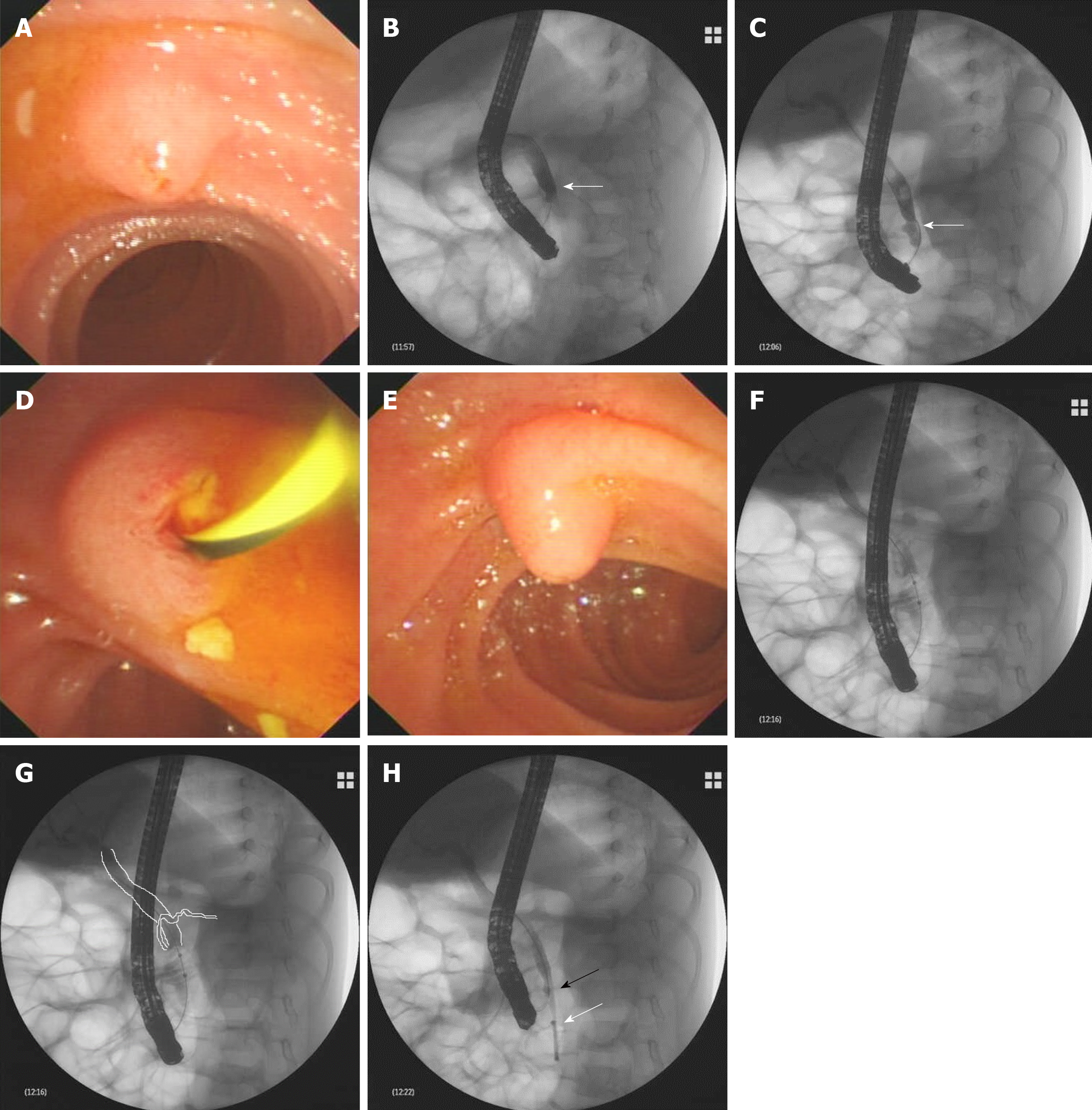
Figure 1 Results of endoscopic retrograde cholangiopancreatography.
A: During endoscopic retrograde cholangiopancreatography, a hemispheric papilla with a villus-like opening resembling the major papilla was seen. B-E: Successful cannulation of the papilla and a dilated common bile duct (CBD) detected by X-ray after injection of a contrast agent (B). Multiple small biliary stones were discharged from the papilla after minor endoscopic sphincterotomy (D). Beneath the papilla, the real major papilla was detected (E). After cannulation of the major papilla, the CBD was observed again, which was dilated with multiple filling defects (C), and at the level of the middle–lower CBD, narrowing was observed (arrow); F: During removal of the biliary stones with an endoscopic balloon, unexpectedly, the dorsal pancreatic duct was revealed at the level of the middle–lower part of the CBD; G: Schematic representation of the image of F; H: After clearance of the CBD with the balloon, an 8.5 Fr 4-cm pigtail plastic pancreatic stent was placed in the biliary duct through the major papilla (white arrow); the minor papilla was cannulated again; and the guidewire was advanced into the CBD (black arrow).
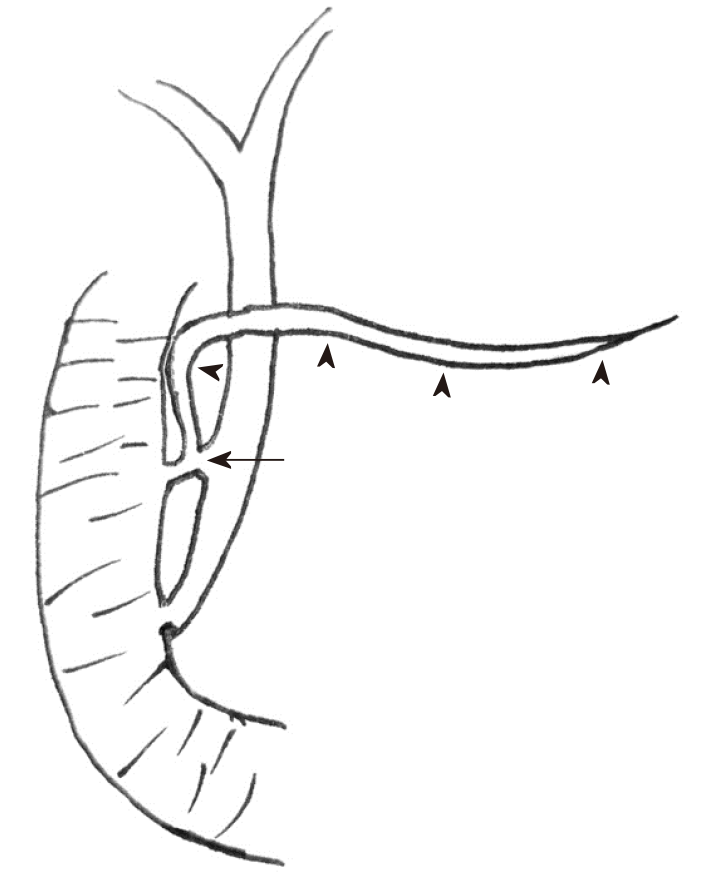
Figure 2 Schematic representation of the pancreaticobiliary system.
This child had three anomalies: Pancreaticobiliary maljunction, pancreas divisum (arrowheads indicating dorsal pancreatic duct), and abnormal communication between the common bile duct and dorsal pancreatic duct (arrow).
- Citation: Cui GX, Huang HT, Yang JF, Zhang XF. Rare variant of pancreaticobiliary maljunction associated with pancreas divisum in a child diagnosed and treated by endoscopic retrograde cholangiopancreatography: A case report. World J Clin Cases 2019; 7(9): 1073-1079
- URL: https://www.wjgnet.com/2307-8960/full/v7/i9/1073.htm
- DOI: https://dx.doi.org/10.12998/wjcc.v7.i9.1073


