Copyright
©The Author(s) 2019.
World J Clin Cases. Apr 6, 2019; 7(7): 809-818
Published online Apr 6, 2019. doi: 10.12998/wjcc.v7.i7.809
Published online Apr 6, 2019. doi: 10.12998/wjcc.v7.i7.809
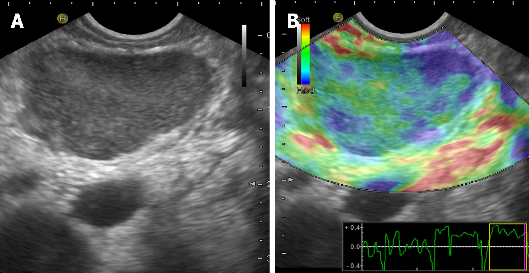
Figure 1 Nodular hypoechoic lesion of the pancreas.
A, B: Nodular hypoechoic lesion of the pancreas showing a mixed pattern (soft versus hard as red and yellow, green and blue colors, respectively) at endoscopic ultrasound elastography.
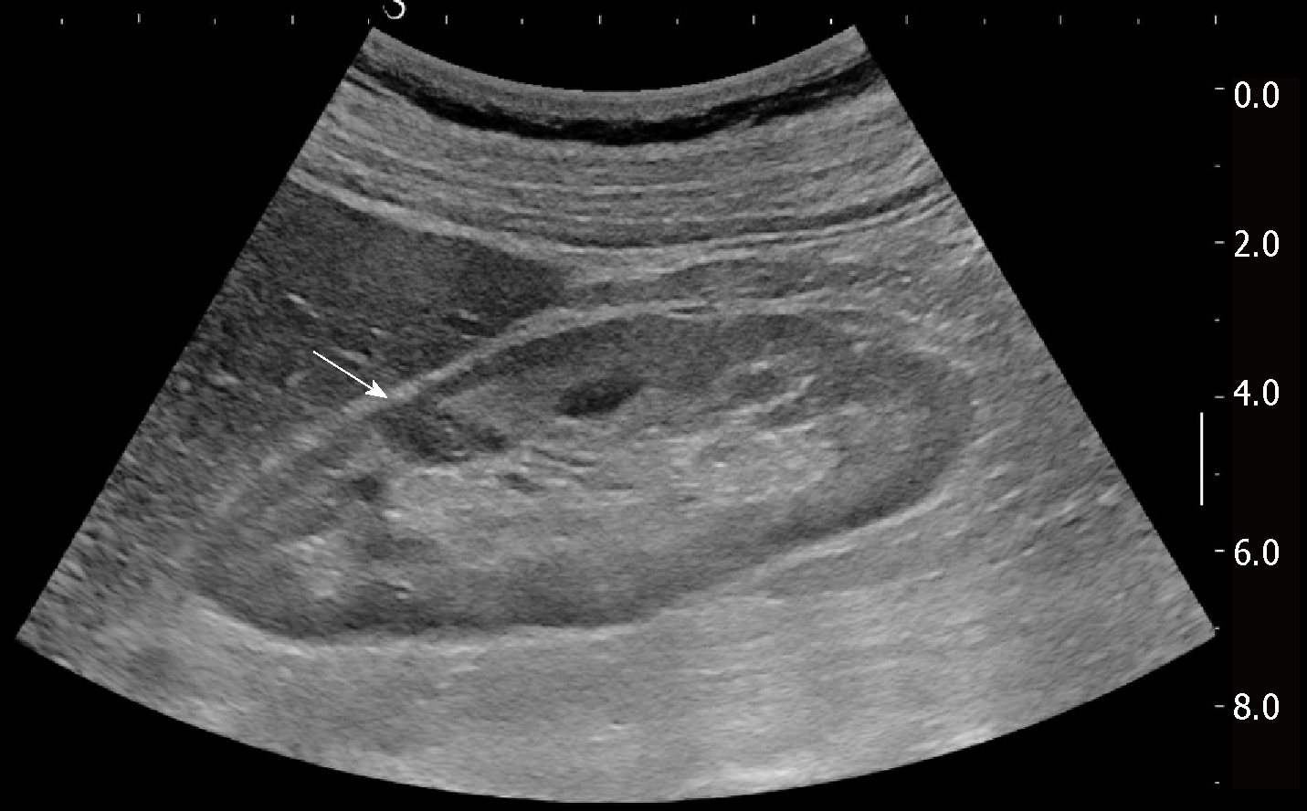
Figure 2 Hypoechoic lesion of the kidney that was revealed as focal nodule from sarcoidosis (arrow).
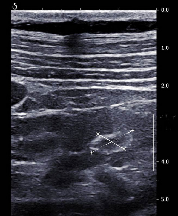
Figure 3 Ring-like echogenic pattern determined by a sarcoid lesion of the renal parenchyma (markers).
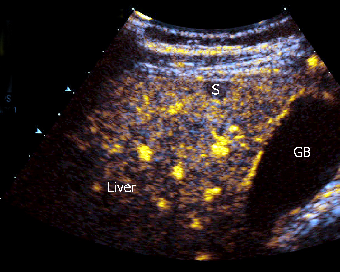
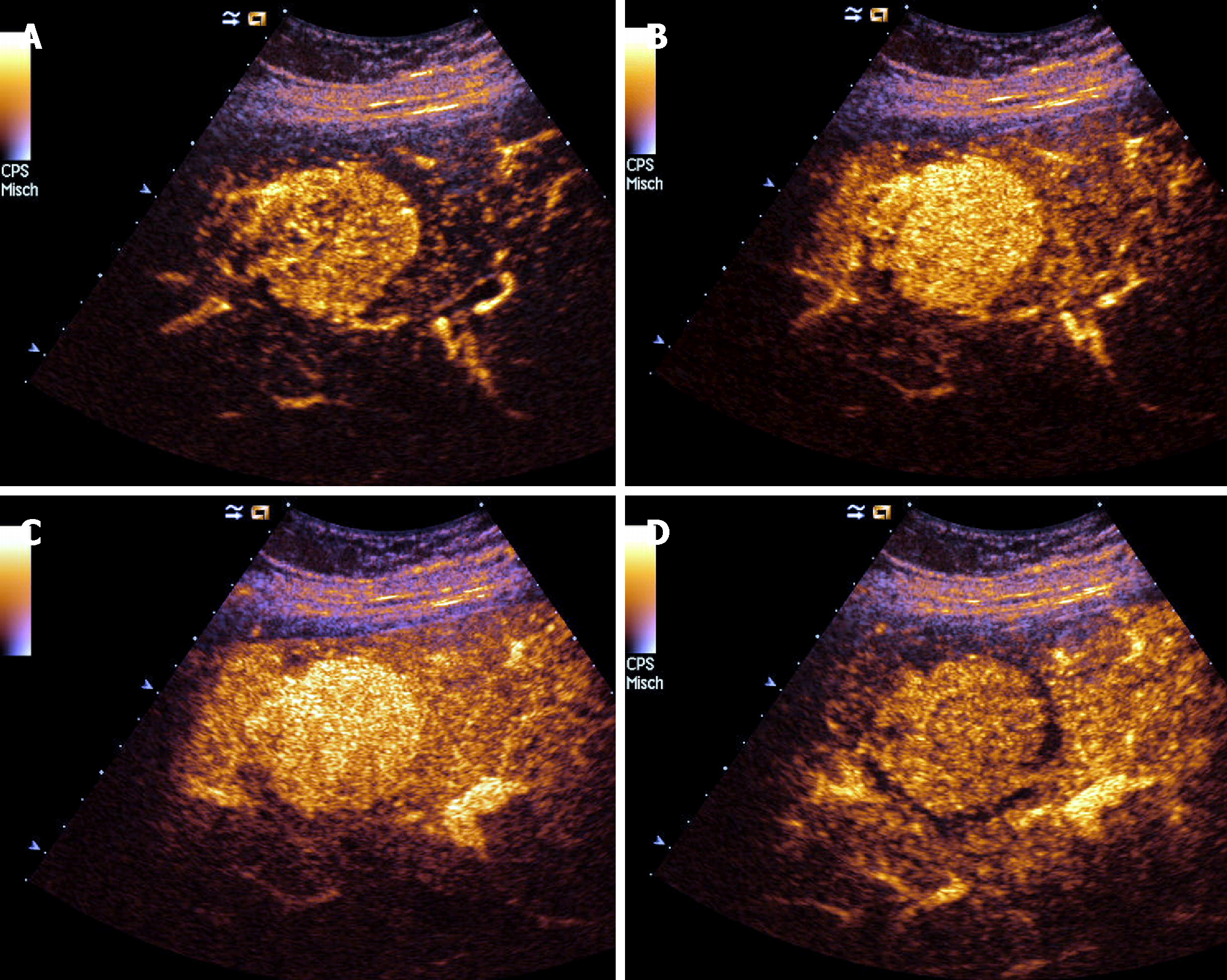
Figure 4 Progressive hypoenhancement in the arterial and portal-venous late phases, respectively, of a nodular sarcoid lesion of the liver.
Figure 5 A rare case of hyperenhancing lesion in arterial, portal-venous and late phase.
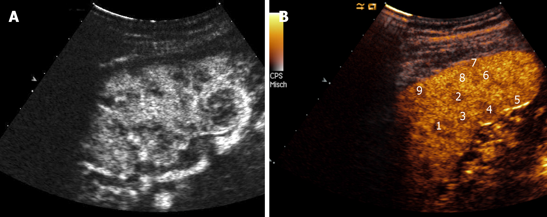
Figure 6 Two different cases of hypoenhancing nodules from sarcoidosis of the spleen.
- Citation: Tana C, Schiavone C, Ticinesi A, Ricci F, Giamberardino MA, Cipollone F, Silingardi M, Meschi T, Dietrich CF. Ultrasound imaging of abdominal sarcoidosis: State of the art. World J Clin Cases 2019; 7(7): 809-818
- URL: https://www.wjgnet.com/2307-8960/full/v7/i7/809.htm
- DOI: https://dx.doi.org/10.12998/wjcc.v7.i7.809


