Copyright
©The Author(s) 2019.
World J Clin Cases. Mar 26, 2019; 7(6): 805-808
Published online Mar 26, 2019. doi: 10.12998/wjcc.v7.i6.805
Published online Mar 26, 2019. doi: 10.12998/wjcc.v7.i6.805
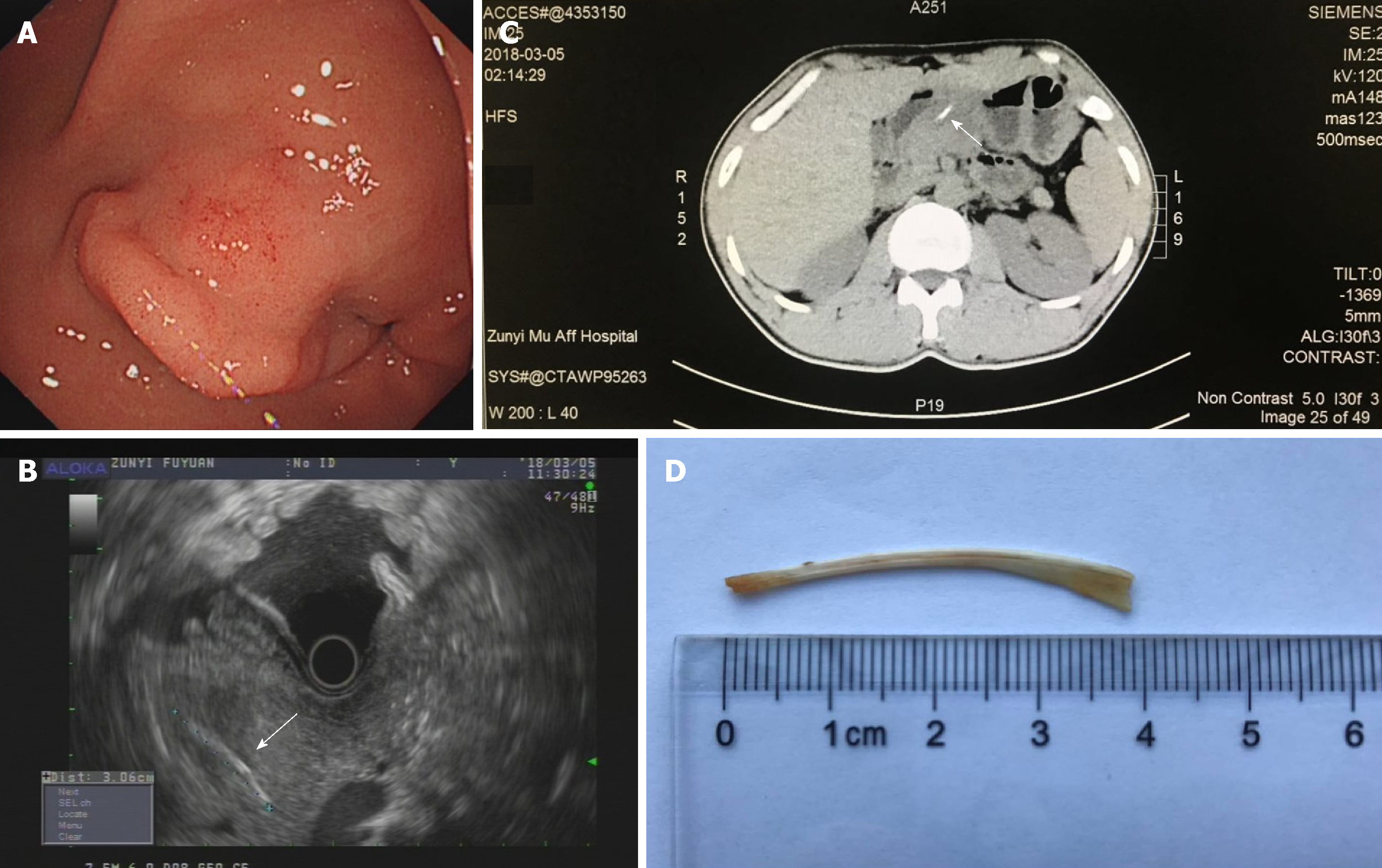
Figure 1 Images of the patient.
A: Upper endoscopy revealed an irregular submucosal tumor on the front wall of the gastric antrum; B: Endoscopic ultrasonography showed abnormal and irregular thickening of the stomach wall and an approximately 3.5-cm linear and hyperechoic lesion protruding through the thickened stomach wall and the pancreatic body (arrow); C: Computed tomography image showing a thin, linear, hyperdense structure (arrow) along the stomach wall and pancreatic body; D: Photograph of the fish bone after removal.
- Citation: Xie R, Tuo BG, Wu HC. Unexplained abdominal pain due to a fish bone penetrating the gastric antrum and migrating into the neck of the pancreas: A case report. World J Clin Cases 2019; 7(6): 805-808
- URL: https://www.wjgnet.com/2307-8960/full/v7/i6/805.htm
- DOI: https://dx.doi.org/10.12998/wjcc.v7.i6.805


