Copyright
©The Author(s) 2019.
World J Clin Cases. Mar 26, 2019; 7(6): 792-797
Published online Mar 26, 2019. doi: 10.12998/wjcc.v7.i6.792
Published online Mar 26, 2019. doi: 10.12998/wjcc.v7.i6.792
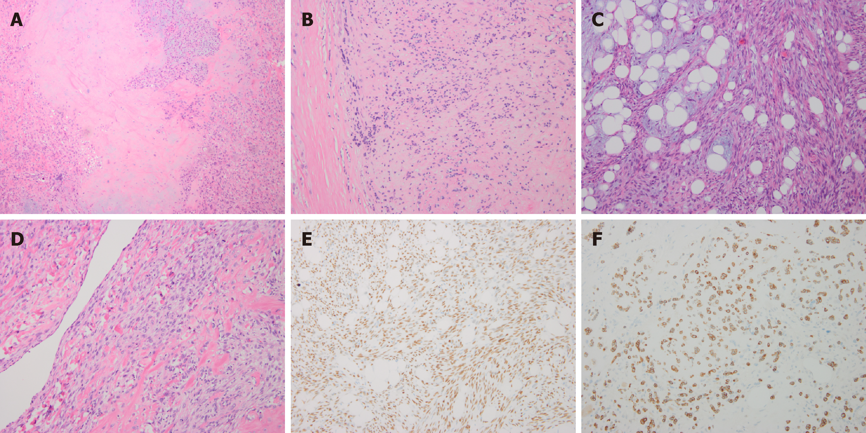
Figure 1 Histopathology features of submandibular gland carcinoma ex pleomorphic adenoma.
A: Myoxid chondriod matrix; B: Carcinoma ex pleomorphic adenoma; C: Spindle cell carcinoma ingredient; D: Osteosarcoma ingredient; E: Immunohistochemical (IHC) analysis revealed P63 nuclear positivity; F: IHC analysis revealed PCK positivity.
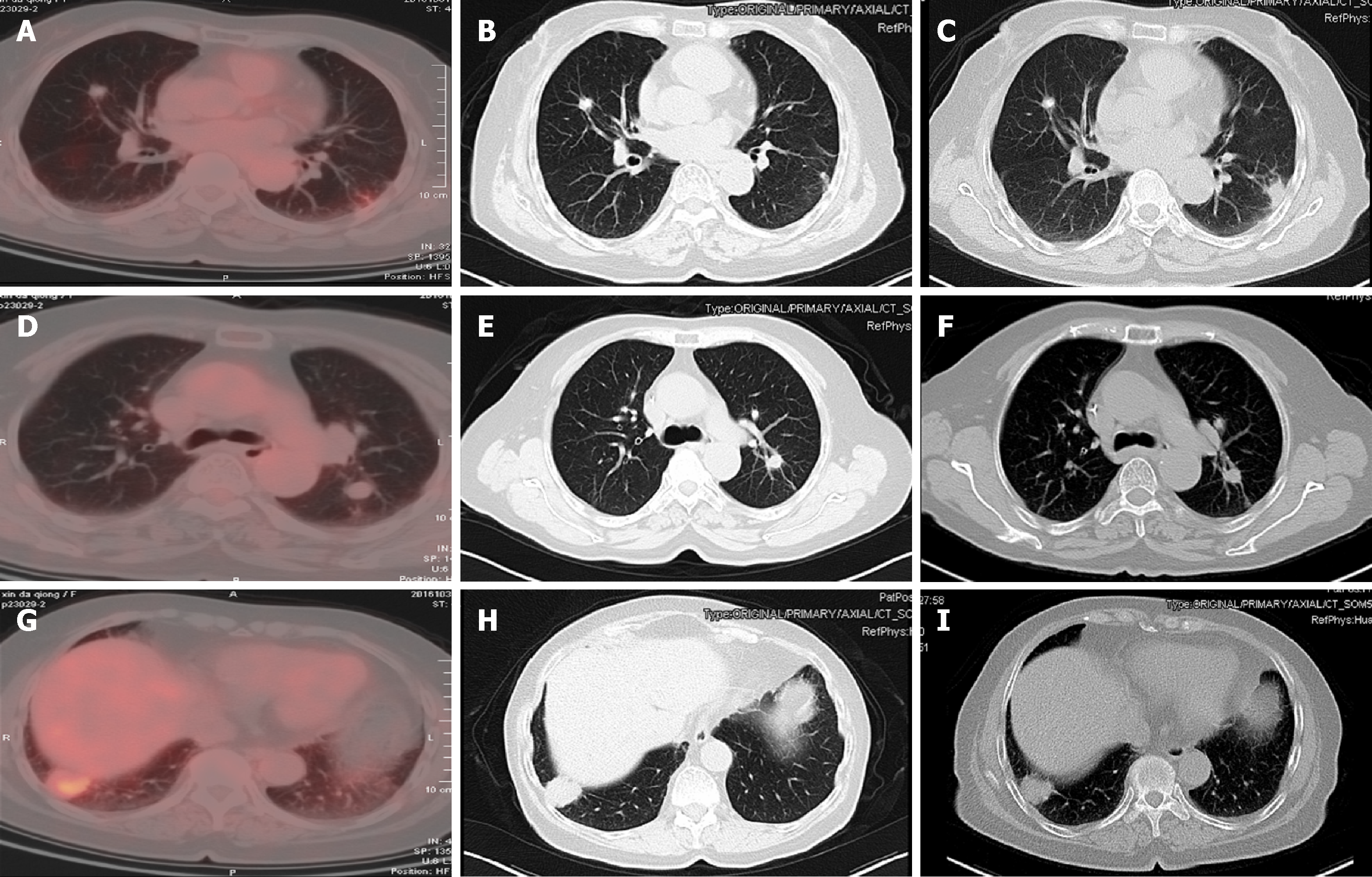
Figure 2 Positron emission tomography-computed tomography and chest enhanced computed tomography images.
The lung metastasis was controlled, and the largest nodule located in lower lobe of the right lung was persistently about 2.5 cm × 2.7 cm in November 2016 (A-C), January 2017 (D-F), and March 2017 (G-I).
- Citation: Chen ZY, Zhang Y, Tu Y, Zhao W, Li M. Effective chemotherapy for submandibular gland carcinoma ex pleomorphic adenoma with lung metastasis after radiotherapy: A case report. World J Clin Cases 2019; 7(6): 792-797
- URL: https://www.wjgnet.com/2307-8960/full/v7/i6/792.htm
- DOI: https://dx.doi.org/10.12998/wjcc.v7.i6.792


