Copyright
©The Author(s) 2019.
World J Clin Cases. Mar 26, 2019; 7(6): 778-784
Published online Mar 26, 2019. doi: 10.12998/wjcc.v7.i6.778
Published online Mar 26, 2019. doi: 10.12998/wjcc.v7.i6.778
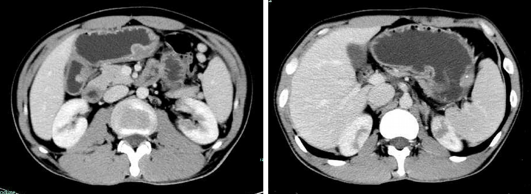
Figure 1 Abdominal computed tomography scan with contrast showed gastric wall thickening and multiple polyps with spotty calcification; progressive enhancement, suggestive of mesenchymal tumors.
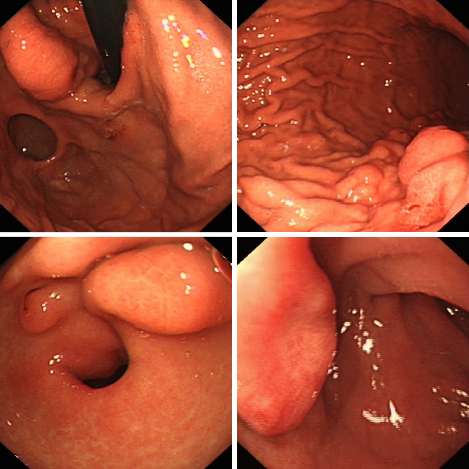
Figure 2 Upper endoscopy revealed multiple submucosal nodules in the antrum, the duodenal bulb, and the gastric body extending to the cardia.
Some nodules had superficial congestion and hemorrhagic spots.
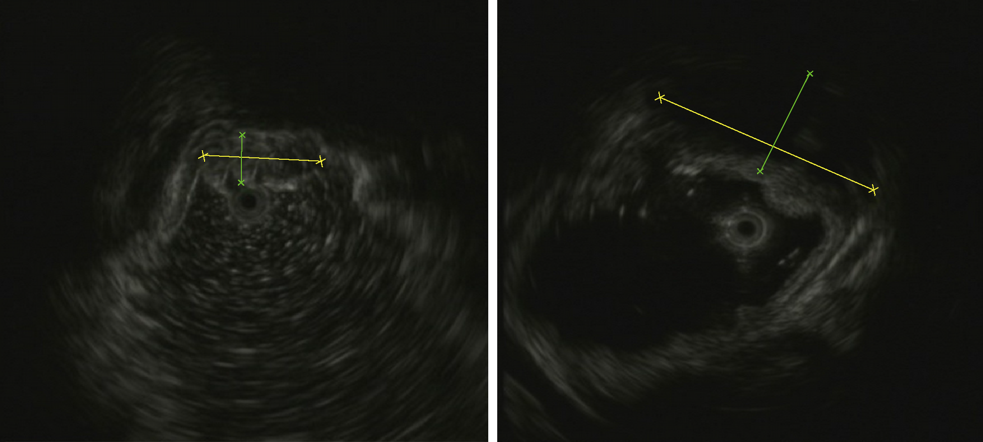
Figure 3 Endoscopic ultrasound showed heterogeneous mixed hypoechoic and iso-echoic lesions in the submucosa and the muscularis propria with hyperechoic spots.
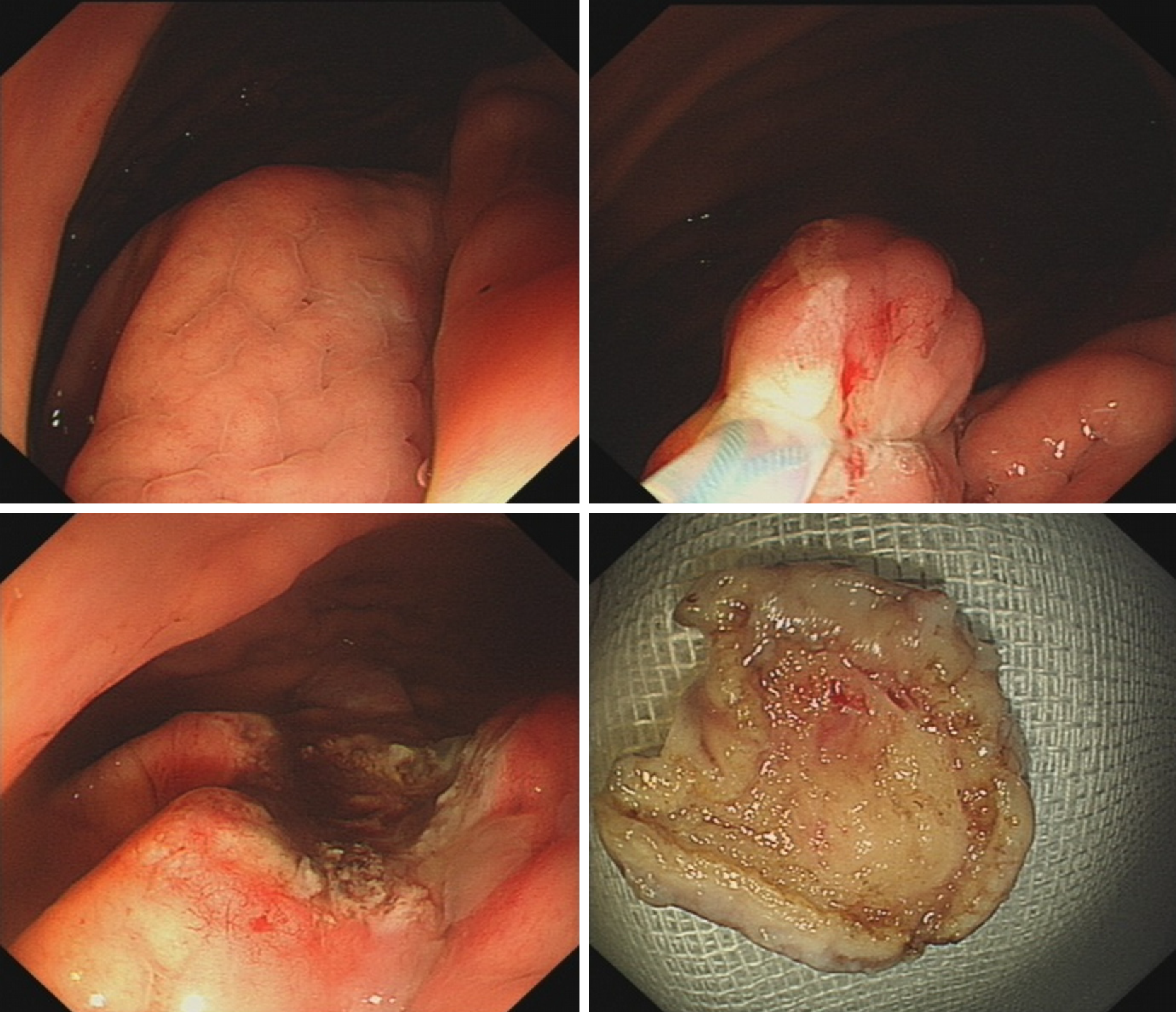
Figure 4 Diagnostic endoscopic snare resection.
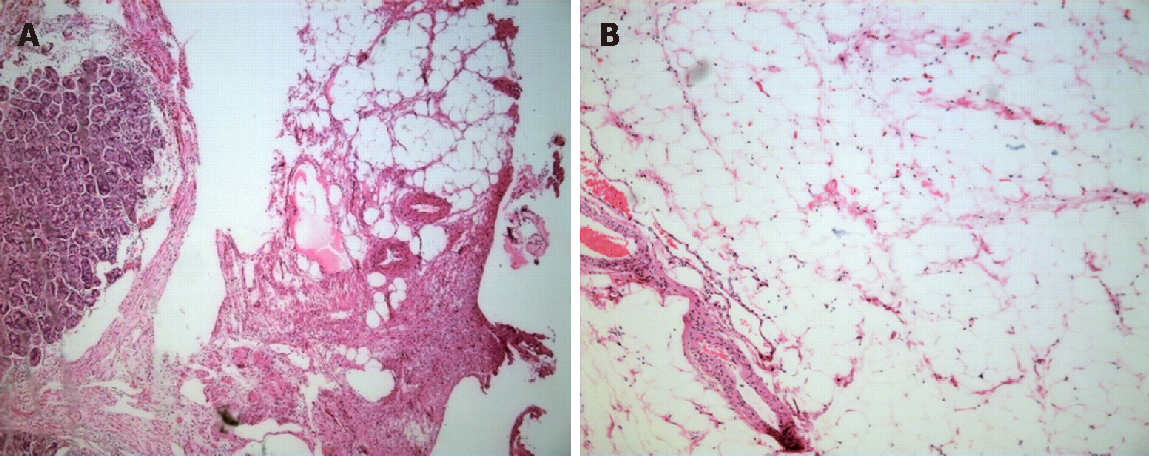
Figure 5 Histological examinations showed a mass consisting of proliferated vascular tissues and mature adipose tissue.
A: HE stain, lower magnification, × 50; B: Higher magnification, × 100. HE: Hematoxylin and eosin.
- Citation: Lou XH, Chen WG, Ning LG, Chen HT, Xu GQ. Multiple gastric angiolipomas: A case report. World J Clin Cases 2019; 7(6): 778-784
- URL: https://www.wjgnet.com/2307-8960/full/v7/i6/778.htm
- DOI: https://dx.doi.org/10.12998/wjcc.v7.i6.778


