Copyright
©The Author(s) 2019.
World J Clin Cases. Mar 26, 2019; 7(6): 742-752
Published online Mar 26, 2019. doi: 10.12998/wjcc.v7.i6.742
Published online Mar 26, 2019. doi: 10.12998/wjcc.v7.i6.742
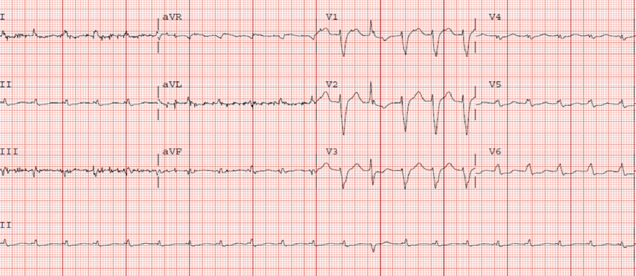
Figure 1 Electrocardiogram – Left bundle branch block with low voltage in limb leads.
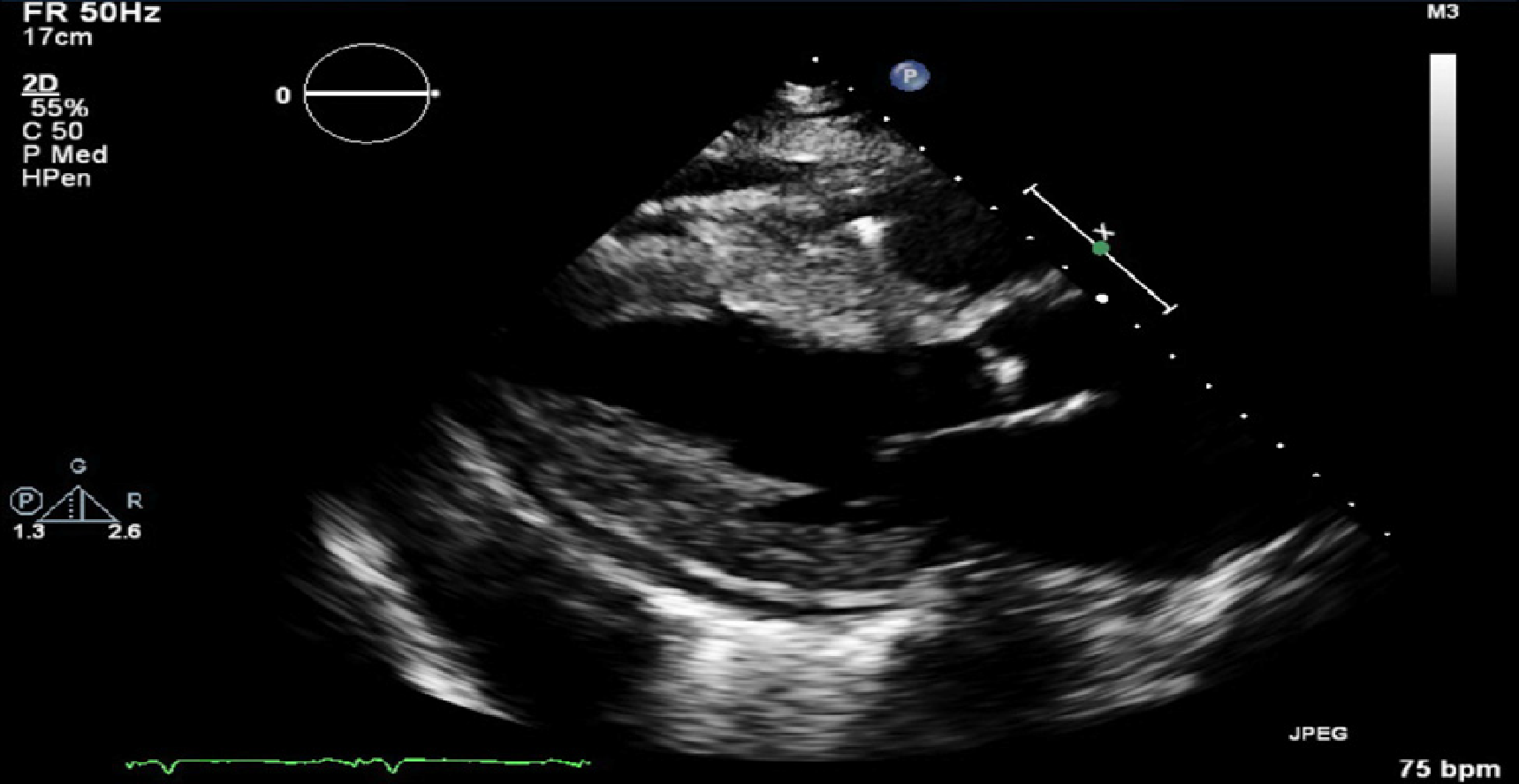
Figure 2 Parasternal long axis echocardiographic view demonstrating severe concentric left ventricular hypertrophy with small pericardial effusion.
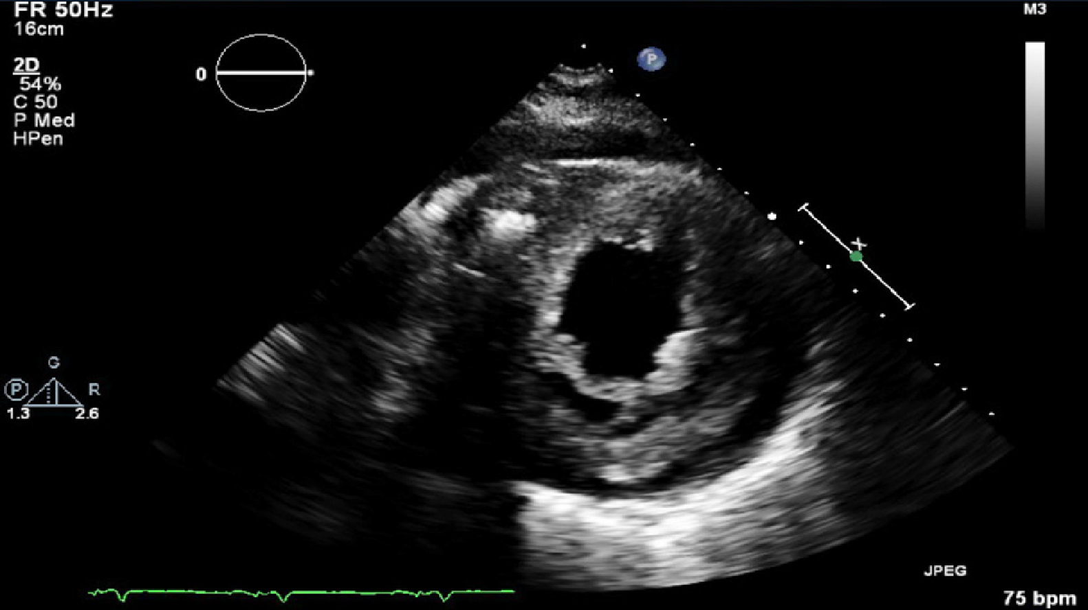
Figure 3 Short axis echocardiographic view demonstrating severe concentric left ventricular hypertrophy with small pericardial effusion.
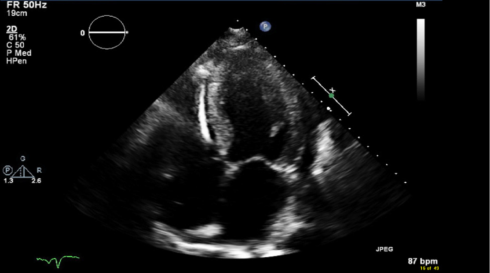
Figure 4 Four chamber echocardiographic view demonstrating bi atrial enlargement, left ventricular hypertrophy, small pericardial effusion and implantable cardioverter-defibrillator device.
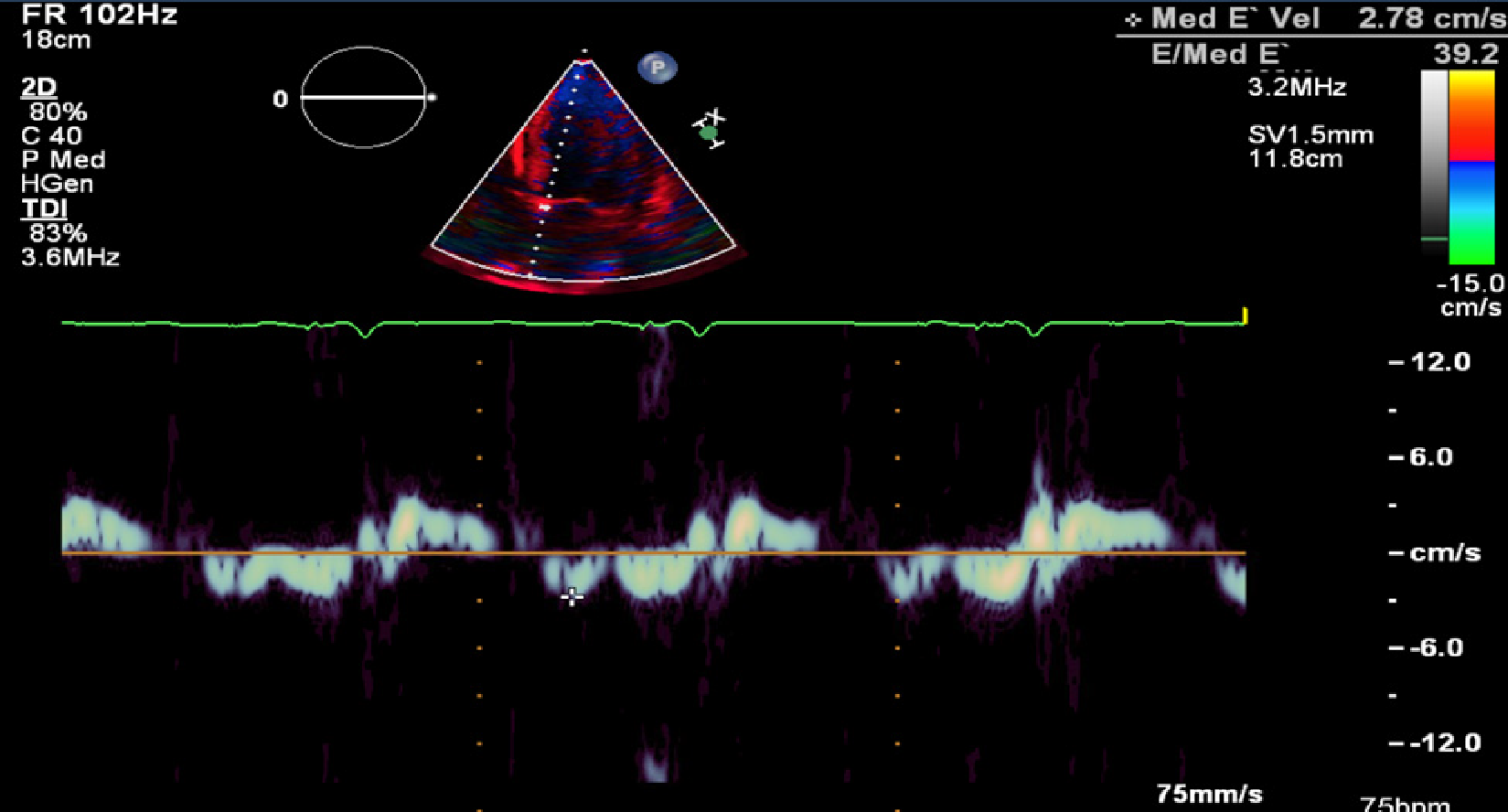
Figure 5 Doppler demonstrating restrictive physiology with medial E’ velocity of 2.
78 cm/s and E/Med E’ 39.2.
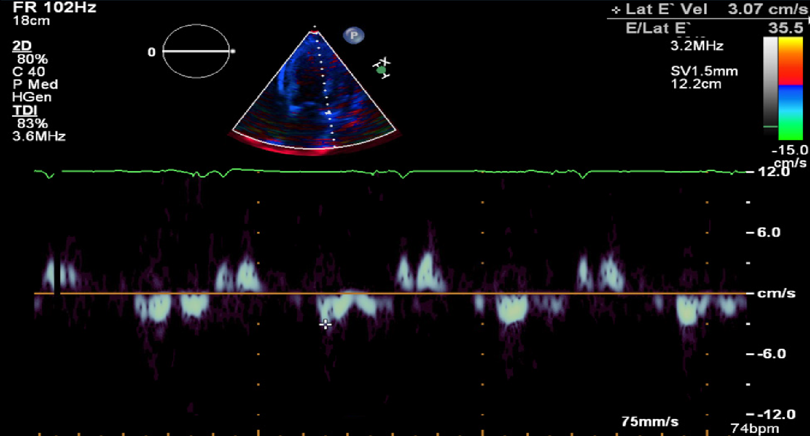
Figure 6 Doppler demonstrating restrictive physiology with lateral E’ velocity of 3.
07cm/s and E/Lat E’ 35.5.
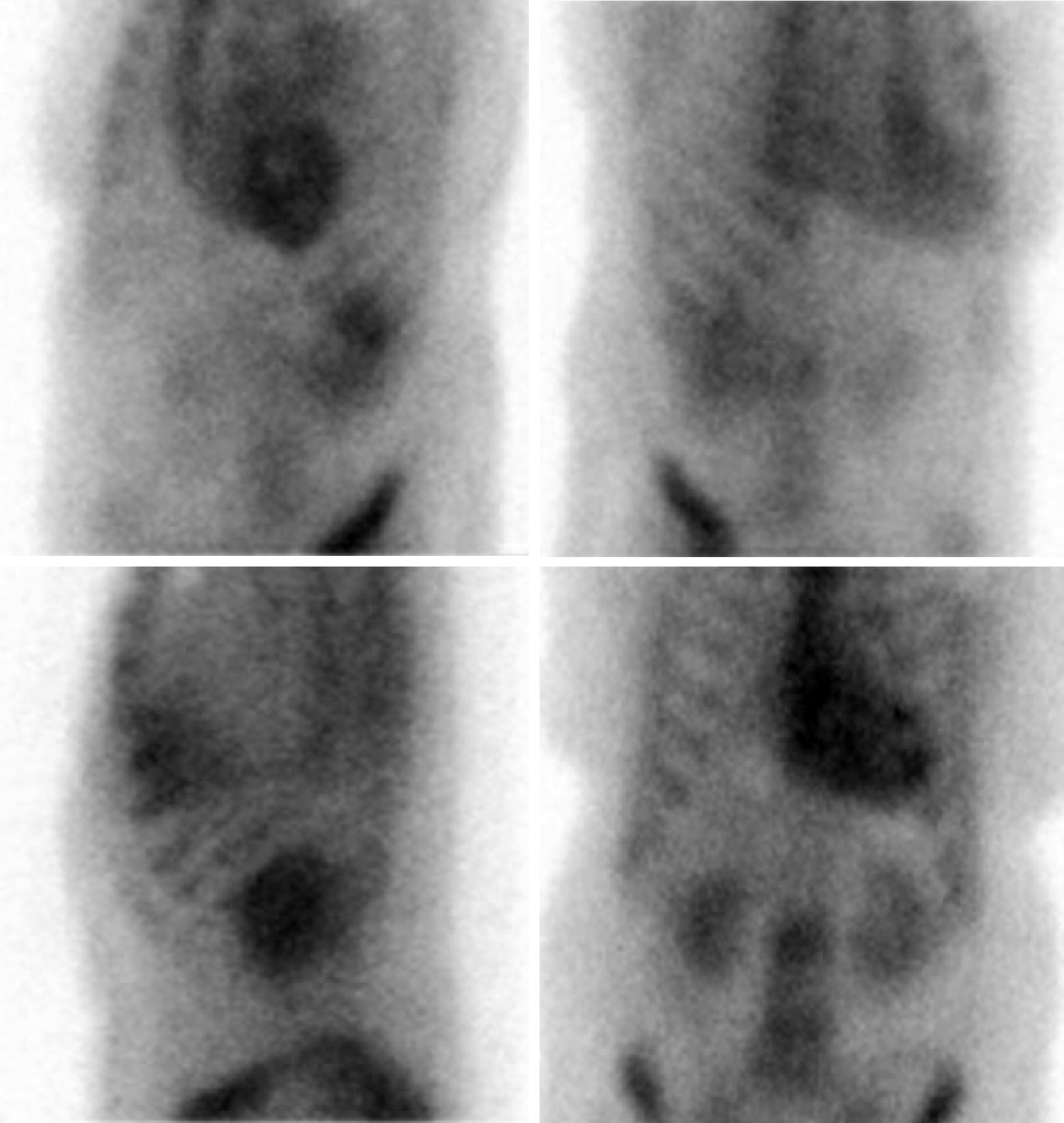
Figure 7 Planar scintigraphy using 99Technetium pyrophosphate for detection of amyloid protein in the heart.
Quantitative analysis of heart retention (done 4 h after injection of 99Technetium pyrophosphate) showed a heart to contralateral (H/CL) ratio of 1.8 (a ratio of > 1.5 is considered to strongly suggest ATTR amyloid deposition).
- Citation: Taiwo AA, Alapati L, Movahed A. Cardiac amyloidosis: A case report and review of literature. World J Clin Cases 2019; 7(6): 742-752
- URL: https://www.wjgnet.com/2307-8960/full/v7/i6/742.htm
- DOI: https://dx.doi.org/10.12998/wjcc.v7.i6.742


