Copyright
©The Author(s) 2019.
World J Clin Cases. Mar 6, 2019; 7(5): 650-655
Published online Mar 6, 2019. doi: 10.12998/wjcc.v7.i5.650
Published online Mar 6, 2019. doi: 10.12998/wjcc.v7.i5.650
Figure 1 Facial features and preoperative intraoral view of the patient.
A: Frontal view of the patient; B: Profile view of the patient; C: Intraoral view. Maxillary and mandibular arches were fused.
Figure 2 Three-dimensional computed tomography images demonstrating bony fusion of bilateral maxillaries to the mandibles.
A: Frontal view; B: Right lateral view; C: Left lateral view.
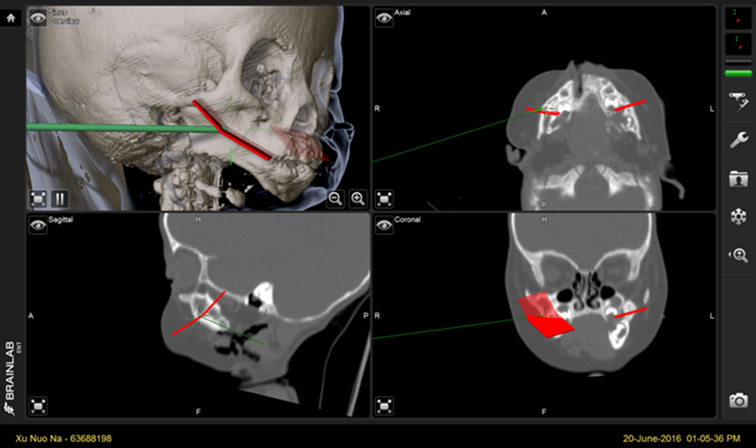
Figure 3 Screenshot of the navigation system.
The pointer indicates arrival at the target site.
- Citation: Lin LQ, Bai SS, Wei M. Application of computer-assisted navigation in treating congenital maxillomandibular syngnathia: A case report. World J Clin Cases 2019; 7(5): 650-655
- URL: https://www.wjgnet.com/2307-8960/full/v7/i5/650.htm
- DOI: https://dx.doi.org/10.12998/wjcc.v7.i5.650


