Copyright
©The Author(s) 2019.
World J Clin Cases. Mar 6, 2019; 7(5): 616-622
Published online Mar 6, 2019. doi: 10.12998/wjcc.v7.i5.616
Published online Mar 6, 2019. doi: 10.12998/wjcc.v7.i5.616
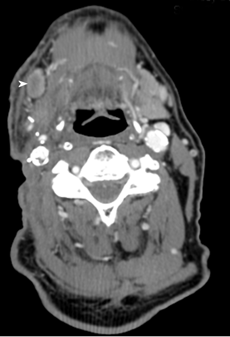
Figure 1 Contrast-enhanced computed tomography scan of neck prior to erlotinib treatment.
Development of right neck focal skin irregularity and right submandibular lymphadenopathy (white arrowhead) consistent with metastases were noted.
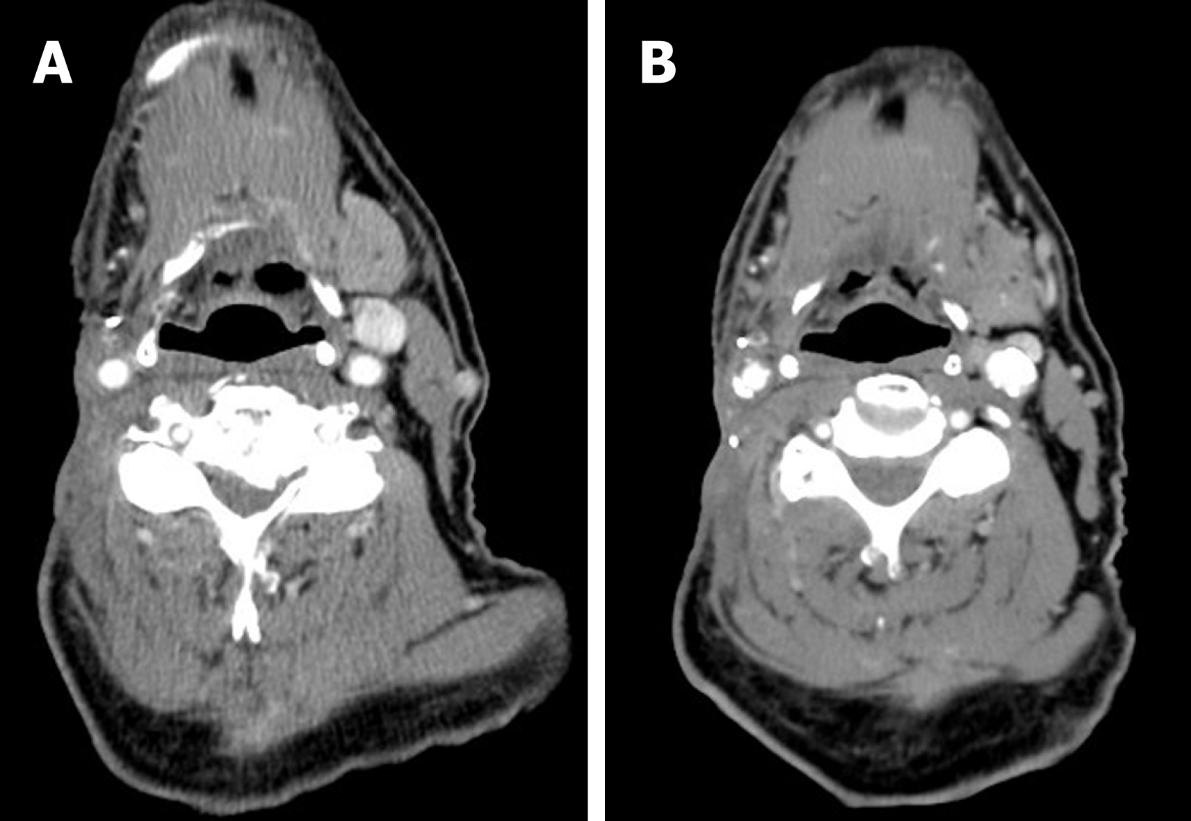
Figure 2 Contrast-enhanced computed tomography scan of neck.
A: After completion of 6-mo erlotinib treatment; B: Seven mo after discontinuation of erlotinib treatment. Both showed resolution of right neck focal skin irregularity and right submandibular lymphadenopathy as shown in Figure 1.
- Citation: Thinn MM, Hsueh CT, Hsueh CT. Sustained complete response to erlotinib in squamous cell carcinoma of the head and neck: A case report. World J Clin Cases 2019; 7(5): 616-622
- URL: https://www.wjgnet.com/2307-8960/full/v7/i5/616.htm
- DOI: https://dx.doi.org/10.12998/wjcc.v7.i5.616


