Copyright
©The Author(s) 2019.
World J Clin Cases. Feb 6, 2019; 7(3): 396-404
Published online Feb 6, 2019. doi: 10.12998/wjcc.v7.i3.396
Published online Feb 6, 2019. doi: 10.12998/wjcc.v7.i3.396
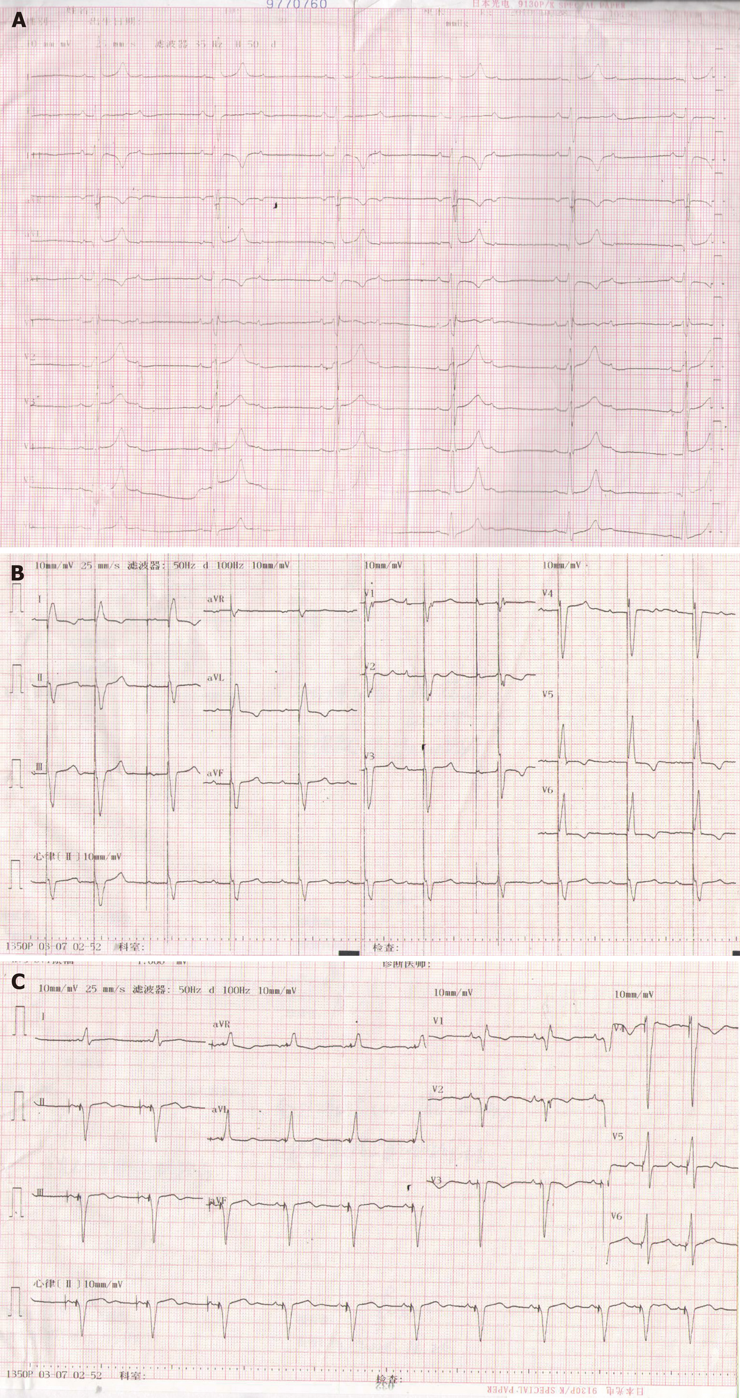
Figure 1 Echocardiography of the patient.
A: Preoperative echocardiography (ECG); B: ECG after DDD (DDD pacing mode, QRS duration 150 ms); C: ECG after cardiac resynchronization therapy (CRT) (CRT pacing mode, QRS duration 120 ms).
Figure 2 Tissue Doppler echocardiography.
A: Synchronicity of mitral valve annulus in tissue Doppler echocardiography after DDD; B: Synchronicity of mitral valve annulus in tissue Doppler echocardiography after cardiac resynchronization therapy.
- Citation: Yu S, Wu Q, Chen BL, An YP, Bu J, Zhou S, Wang YM. Biventricular pacing for treating heart failure in children: A case report and review of the literature. World J Clin Cases 2019; 7(3): 396-404
- URL: https://www.wjgnet.com/2307-8960/full/v7/i3/396.htm
- DOI: https://dx.doi.org/10.12998/wjcc.v7.i3.396


