Copyright
©The Author(s) 2019.
World J Clin Cases. Feb 6, 2019; 7(3): 389-395
Published online Feb 6, 2019. doi: 10.12998/wjcc.v7.i3.389
Published online Feb 6, 2019. doi: 10.12998/wjcc.v7.i3.389
Figure 1 Proband’s pedigree.
Squares indicate men, circles indicate women, black symbols indicate affected individuals, diagonal lines across symbols indicate deceased individuals, and the arrow indicates the proband.
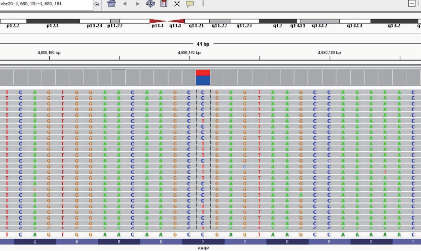
Figure 2 Gene mapping of the proband.
A heterozygous mutation site was found in the 2nd exonic sequence of the PRNP gene (c.305; cytosine to thymine), resulting in a p.P102L amino acid change (valine to leucine).
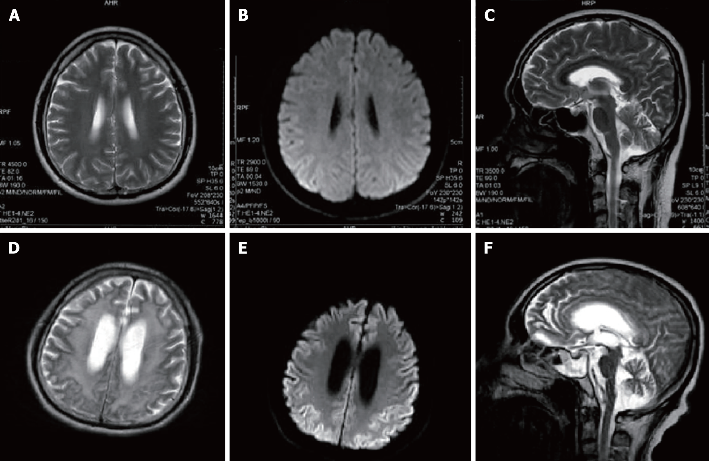
Figure 3 Magnetic resonance imaging scans of the patient’s brain(A, B, D and E: Axial; C and F: Sagittal).
A, B, and C: Brain magnetic resonance images were obtained 2.5 years after symptom onset. There are no obviously abnormal signals in the T2-weighted images (A, B) or diffusion weighted image (DWI) (C); D, E and F: Brain magnetic resonance images were obtained 5.5 years after the onset of symptoms. T2-weighted images (D, F) reveal high signal intensities in the basal ganglia, corona radiata, and paraventricular and semiovale centers; severe brain atrophy; and ventricular dilation. DWI imaging (E) reveals a diffuse symmetrical high signal in the bilateral cerebral cortex.
- Citation: Zhao MM, Feng LS, Hou S, Shen PP, Cui L, Feng JC. Gerstmann-Sträussler-Scheinker disease: A case report. World J Clin Cases 2019; 7(3): 389-395
- URL: https://www.wjgnet.com/2307-8960/full/v7/i3/389.htm
- DOI: https://dx.doi.org/10.12998/wjcc.v7.i3.389


