Copyright
©The Author(s) 2019.
World J Clin Cases. Dec 6, 2019; 7(23): 4163-4171
Published online Dec 6, 2019. doi: 10.12998/wjcc.v7.i23.4163
Published online Dec 6, 2019. doi: 10.12998/wjcc.v7.i23.4163
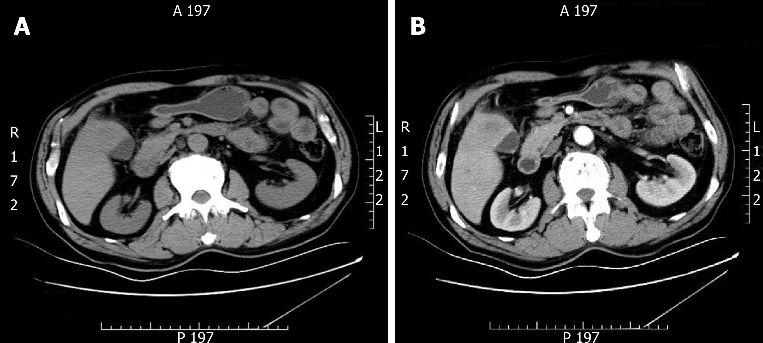
Figure 1 Gallbladder carcinoma infiltrating the adjacent liver, with enlarged retroperitoneal lymph nodes.
A: Computed tomography; B: Contrast enhanced computed tomography.
Figure 2 Gross morphology of a 5 cm × 4 cm × 6 cm tumor seen near the gallbladder neck and liver tissue.
Part of the mass was grey and soft, part of the mass section showing purulent necrosis was visible, and the boundary between the mass and surrounding liver tissue was not clear.
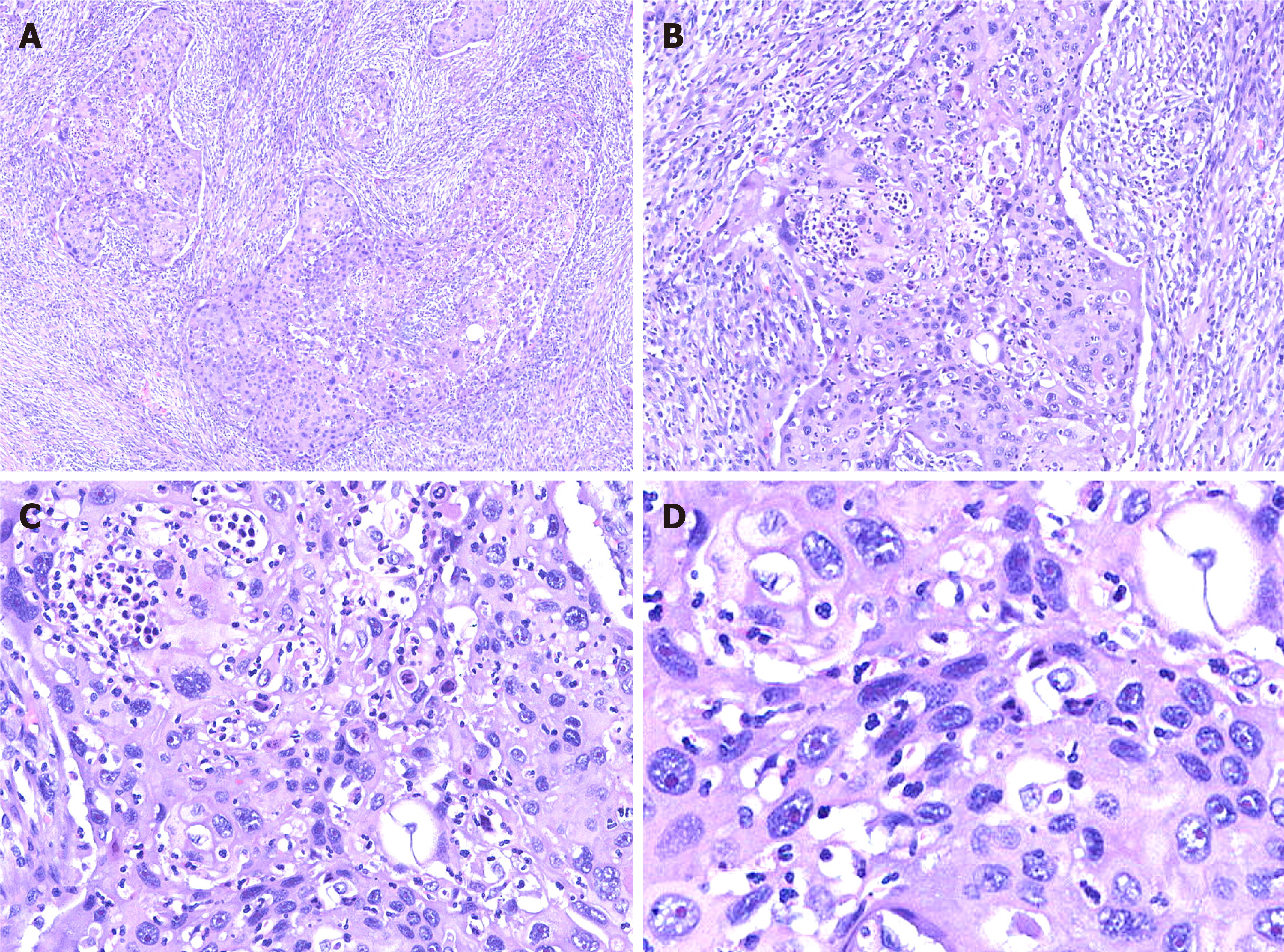
Figure 3 Hematoxylin and eosin staining of tumor cells.
A: Image showing diffuse growth replacing the mucosa and invading the muscularis propria; B: Image showing densely packed tumors formed by squamous cells; C: Image showing squamous cell carcinoma with keratin pearl formation; D: Image showing that tumor cell proliferation is markedly atypical, and karyokinesis is increased.
Figure 4 Immunohistochemical staining shows tumor cells in gallbladder squamous cell carcinoma (×100).
A: AE1/AE3 (+); B: CK5/6 (+); C: CAM5.2 (+); D: CK7 (+); E: CK19 (+); F: P40 (+); G: Hepatocyte (-); H: AFP (-); I: Ki-67 (60%+).
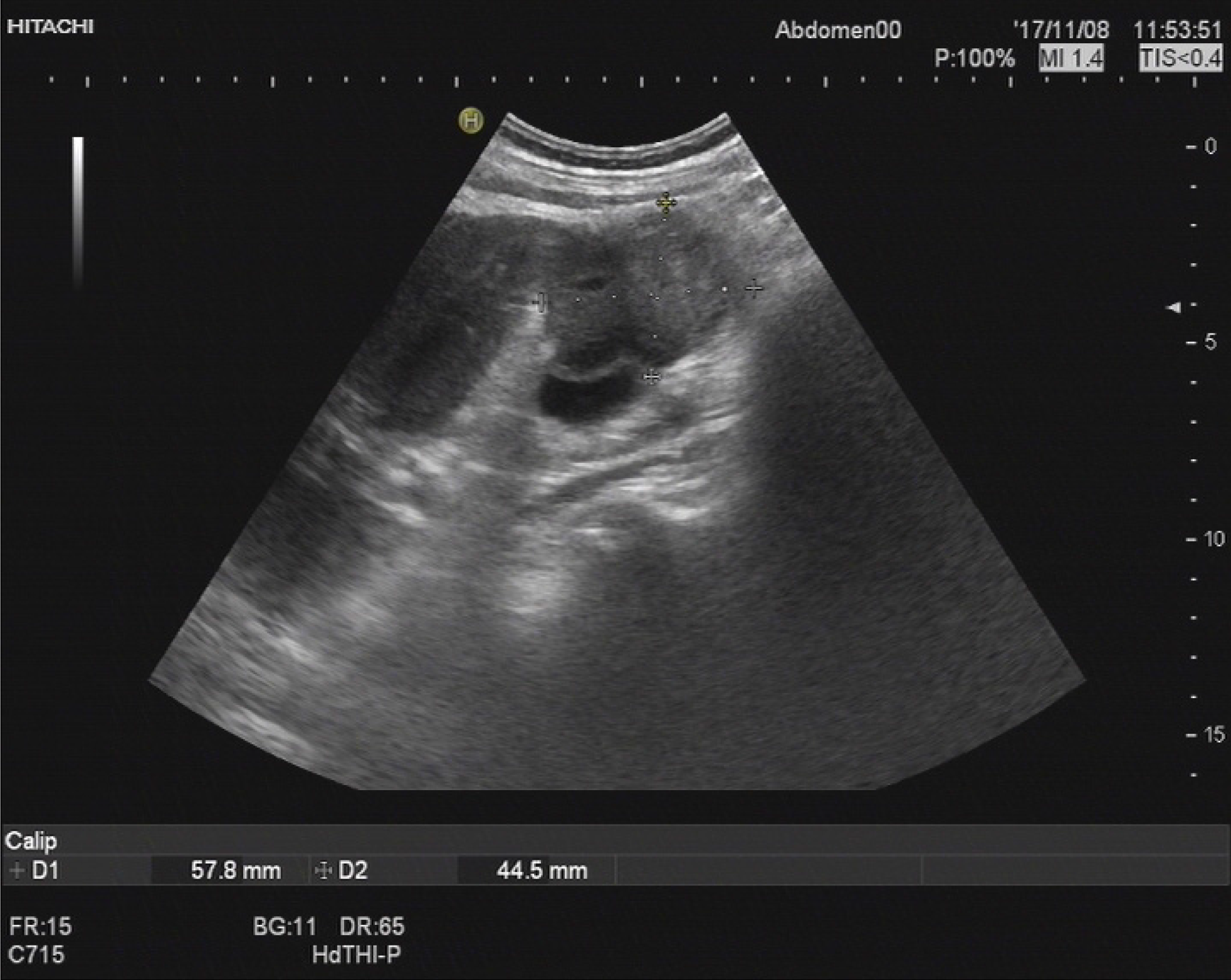
Figure 5 Abdominal ultrasonography revealed a space-occupying lesion in the right posterior aspect of the first hepatic portal region, which was considered as cancer metastasis.
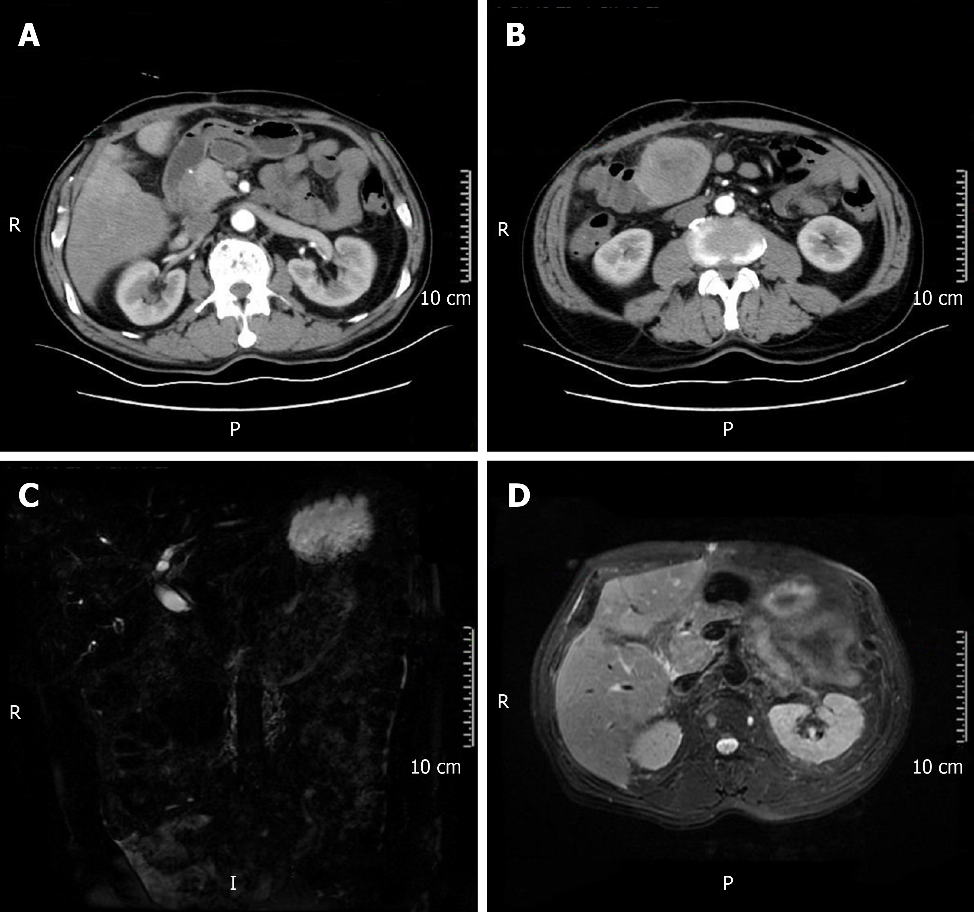
Figure 6 Postoperative changes in gallbladder carcinoma and multiple metastases in the liver and abdominal cavity.
A and B: Computed tomography; C and D: Magnetic resonance cholangiopancreatography.
- Citation: Jin S, Zhang L, Wei YF, Zhang HJ, Wang CY, Zou H, Hu JM, Jiang JF, Pang LJ. Pure squamous cell carcinoma of the gallbladder locally invading the liver and abdominal cavity: A case report and review of the literature. World J Clin Cases 2019; 7(23): 4163-4171
- URL: https://www.wjgnet.com/2307-8960/full/v7/i23/4163.htm
- DOI: https://dx.doi.org/10.12998/wjcc.v7.i23.4163


