Copyright
©The Author(s) 2019.
World J Clin Cases. Sep 26, 2019; 7(18): 2899-2904
Published online Sep 26, 2019. doi: 10.12998/wjcc.v7.i18.2899
Published online Sep 26, 2019. doi: 10.12998/wjcc.v7.i18.2899
Figure 1 Chest computed tomography.
A: Calcification was observed around some nodules; B: A fat-dense shadow could be seen in the anterior mediastinum. There was no enhancement observed by the enhanced computed tomography scan; C: Multiple variably sized fat-dense nodules.
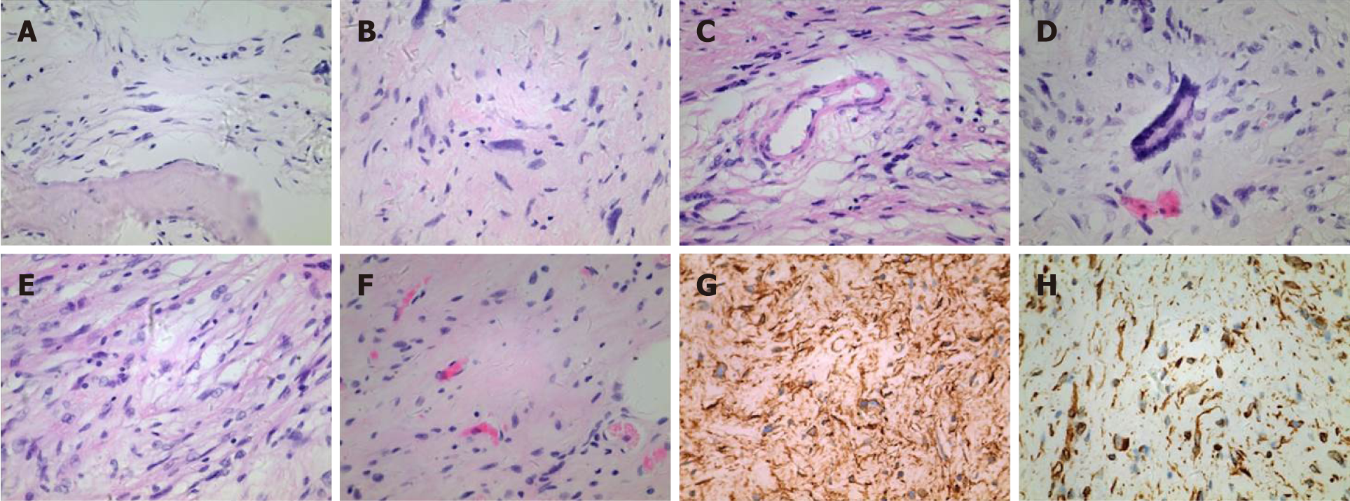
Figure 2 Pathology examination.
A: Intratumoral ossification; B: Atypical nuclei in the tumor; C: A small number of thick-walled blood vessels; D: Floret-like giant cells in the tumor; E: Spindle cells with mild nuclear atypia; F: Thick-walled vessels were observed; G: Immunohistochemical analysis for CD34 (+); H: Immunohistochemical analysis for vimentin (+). Magnification, ×400.
Figure 3 The tumor and cut surface.
A: Poorly defined border between the tumor and pericardium; B: Multiple tumors in the anterior mediastinum; C: Cut surface was grayish yellow.
- Citation: Mao YQ, Liu XY, Han Y. Pleomorphic lipoma in the anterior mediastinum: A case report. World J Clin Cases 2019; 7(18): 2899-2904
- URL: https://www.wjgnet.com/2307-8960/full/v7/i18/2899.htm
- DOI: https://dx.doi.org/10.12998/wjcc.v7.i18.2899


