Copyright
©The Author(s) 2019.
World J Clin Cases. Sep 26, 2019; 7(18): 2879-2887
Published online Sep 26, 2019. doi: 10.12998/wjcc.v7.i18.2879
Published online Sep 26, 2019. doi: 10.12998/wjcc.v7.i18.2879
Figure 1 Preoperative imaging data of the patient.
A and B: Arterial computed tomography; C: Venous CT; D: Magnetic resonance cholangiopancreatography.
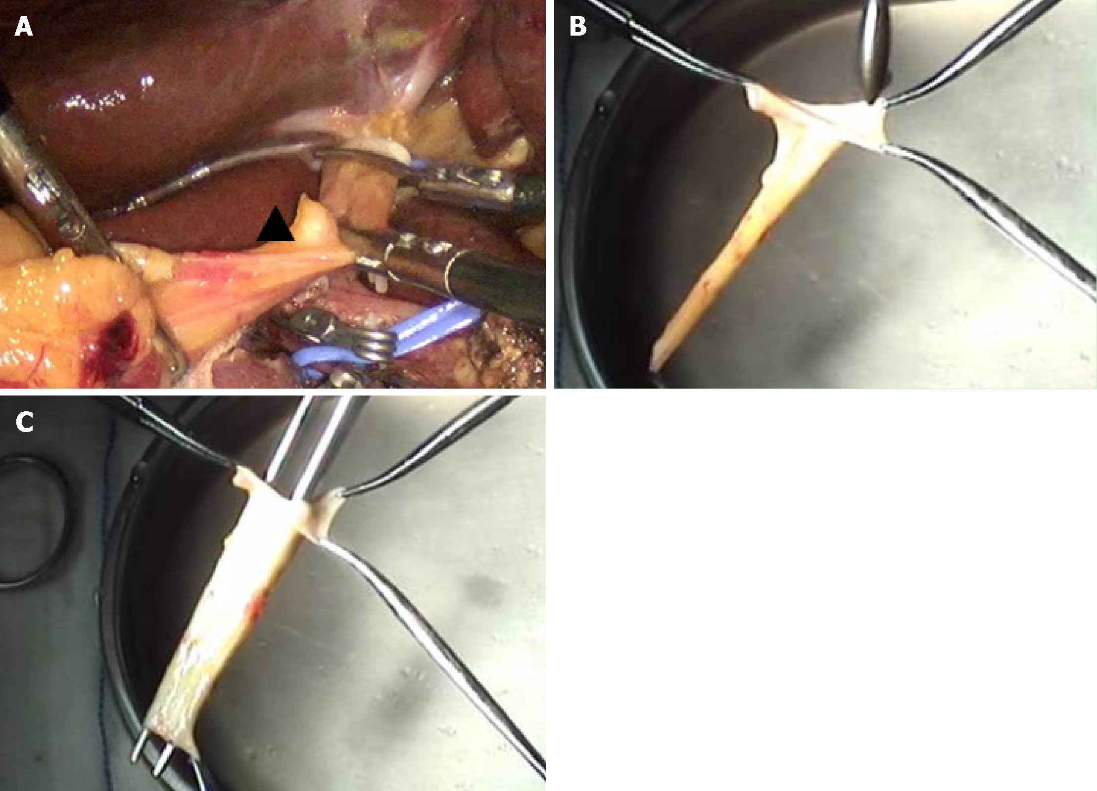
Figure 2 Intraoperative procedure.
A: Removal of autologous hepatic ligamentum teres; B: Dilatation with a bougie and recanalization of the hepatic ligamentum teres; C: Pruning and recanalization of the hepatic ligamentum teres.
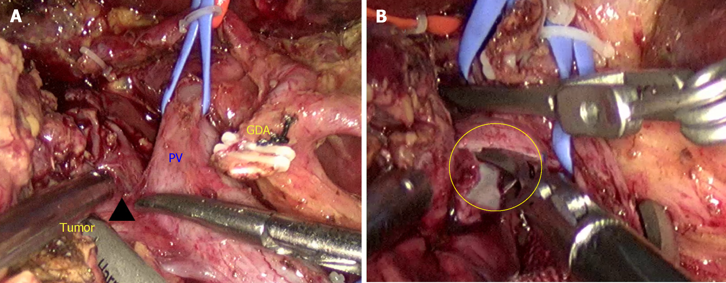
Figure 3 Intraoperative procedure.
A: Part of the right portal vein wall invaded by the tumor was discovered; B: Resection of the portal vein wall invaded by the tumor after blocking the portal vein, and the portal vein defect was about 2.5 cm × 1 cm.
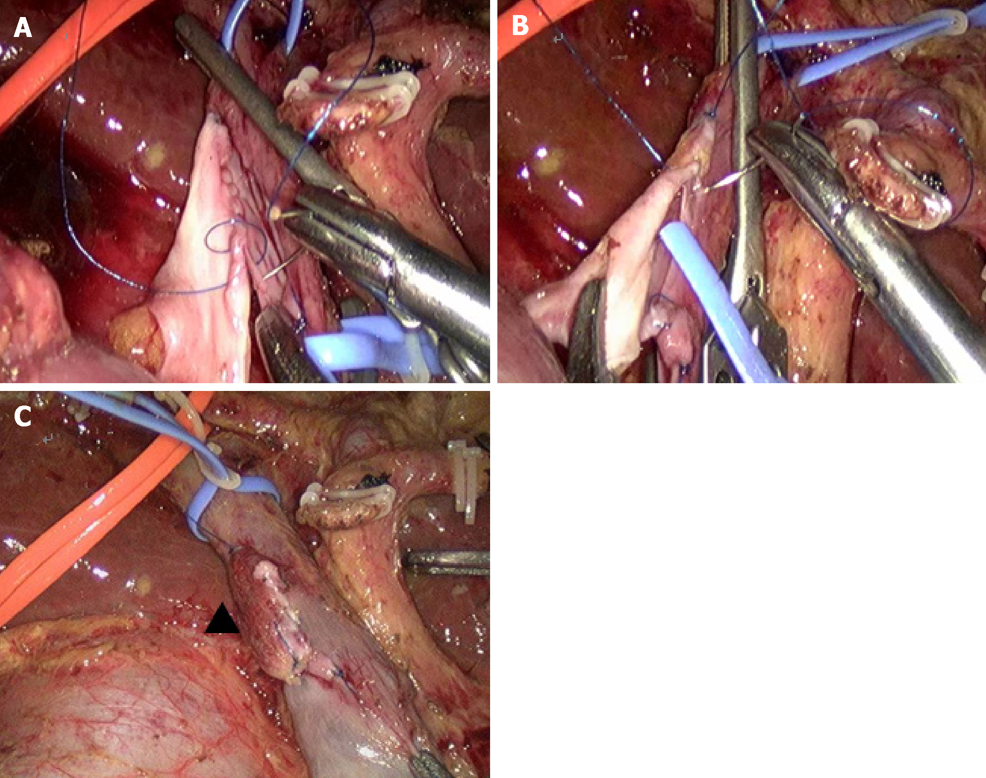
Figure 4 Intraoperative procedure.
A: Repair of the posterior wall of the portal vein; B: Repair of the anterior wall of the portal vein; C: Opening of the portal vein.
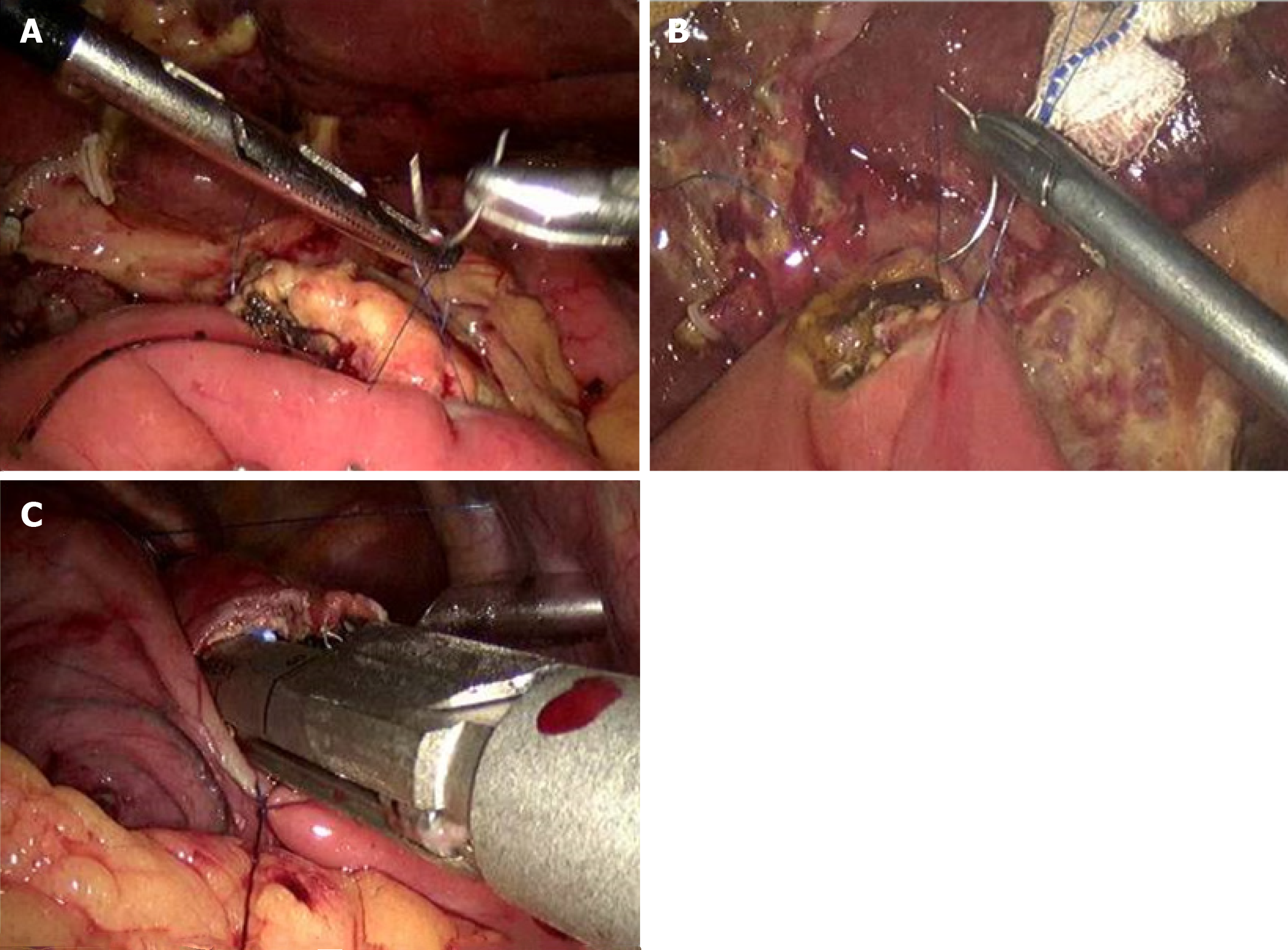
Figure 5 Intraoperative procedure.
A: Pancreaticojejunostomy; B: Cholangiojejunostomy; C: Gastrojejunostomy.
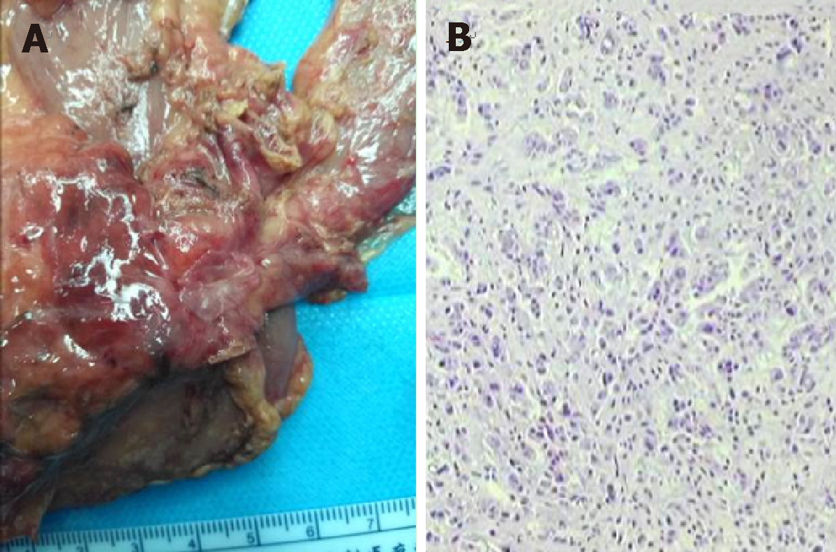
Figure 6 Postoperative and follow-up examination.
A: Posterior view of the specimen; B: Postoperative pathology
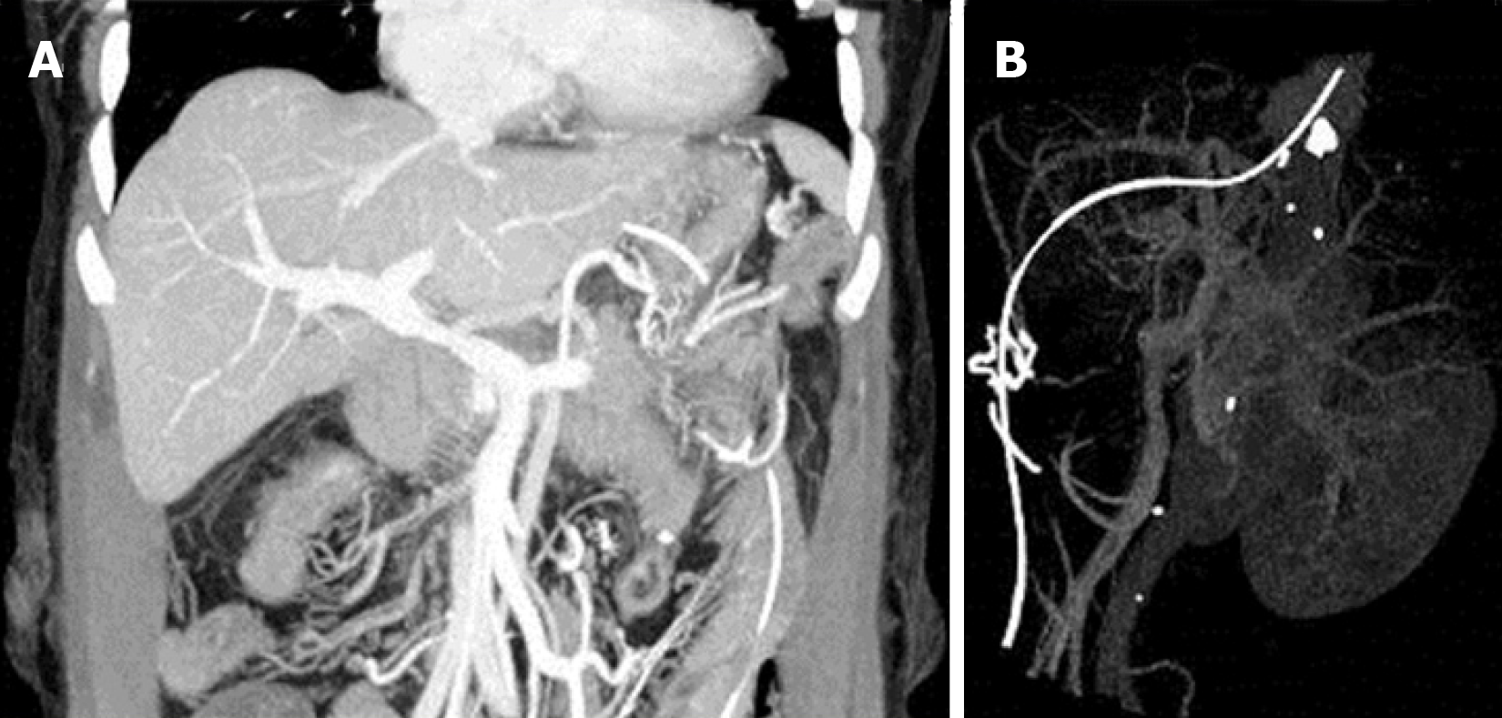
Figure 7 Portal vein computed tomography venography after the operation.
A and B: Portal vein computed tomography taken on July 26
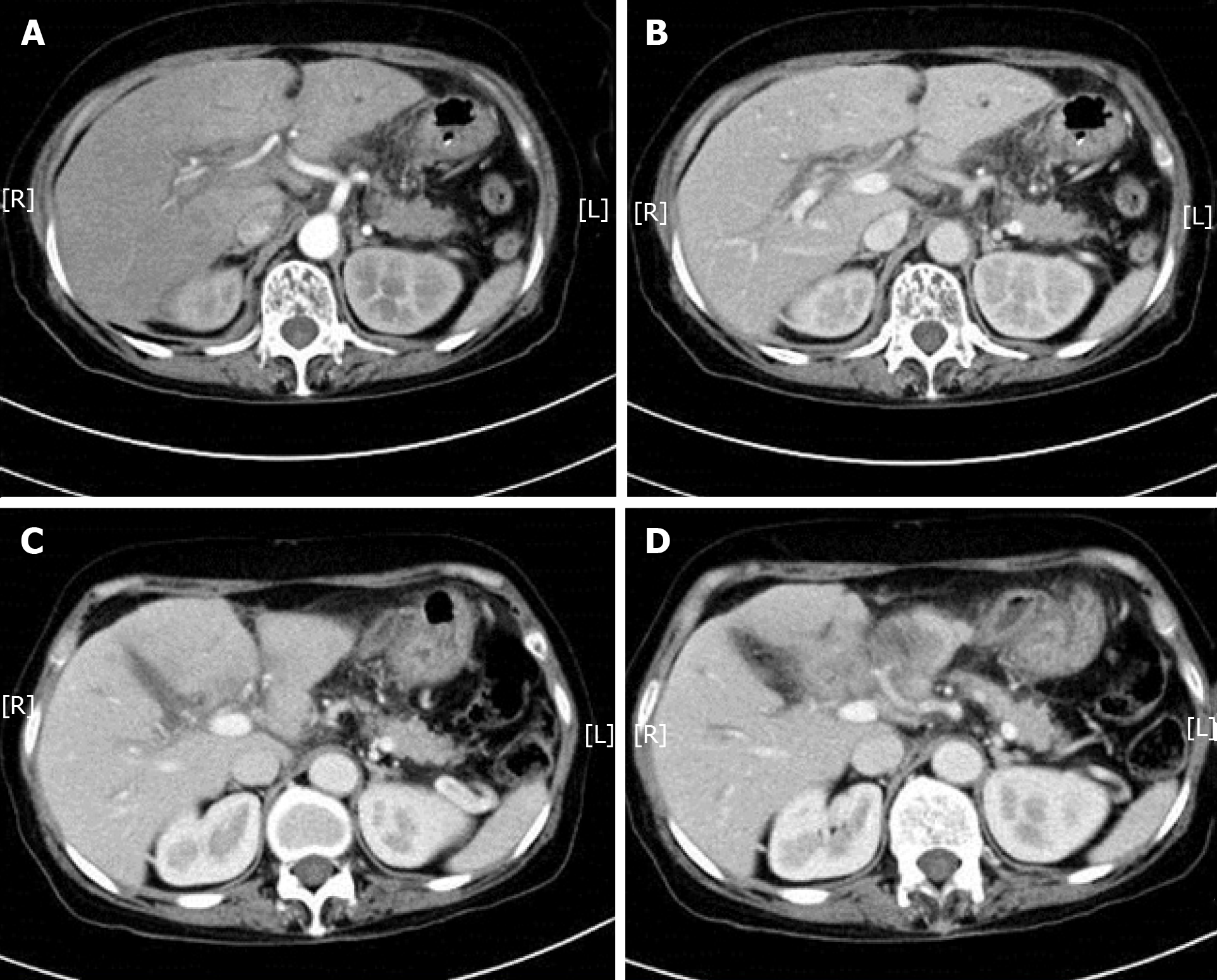
Figure 8 Postoperative and follow-up examination.
A and B: Computed tomography (CT) on July 26; C and D: CT on August 6.
- Citation: Wei Q, Chen QP, Guan QH, Zhu WT. Repair of the portal vein using a hepatic ligamentum teres patch for laparoscopic pancreatoduodenectomy: A case report. World J Clin Cases 2019; 7(18): 2879-2887
- URL: https://www.wjgnet.com/2307-8960/full/v7/i18/2879.htm
- DOI: https://dx.doi.org/10.12998/wjcc.v7.i18.2879


