Copyright
©The Author(s) 2019.
World J Clin Cases. Sep 26, 2019; 7(18): 2843-2850
Published online Sep 26, 2019. doi: 10.12998/wjcc.v7.i18.2843
Published online Sep 26, 2019. doi: 10.12998/wjcc.v7.i18.2843
Figure 1 Preoperative imaging diagnosis.
A: The aortic dissection involved the ascending aorta (yellow arrow); B: The aortic dissection involved the descending thoracic aorta, which extended to the iliac arteries (yellow arrow); C: The aortic dissection involved the aortic arch (yellow arrow).
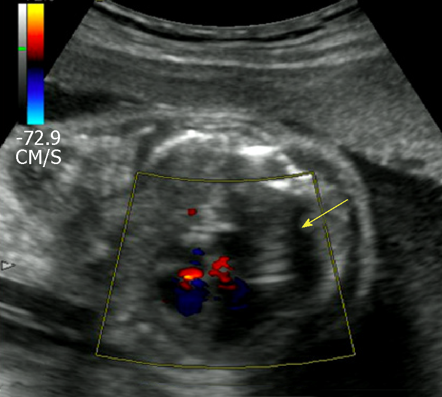
Figure 2 Postoperative ultrasound of the fetus.
The normal heart blood flow of the fetal is shown (yellow arrow).
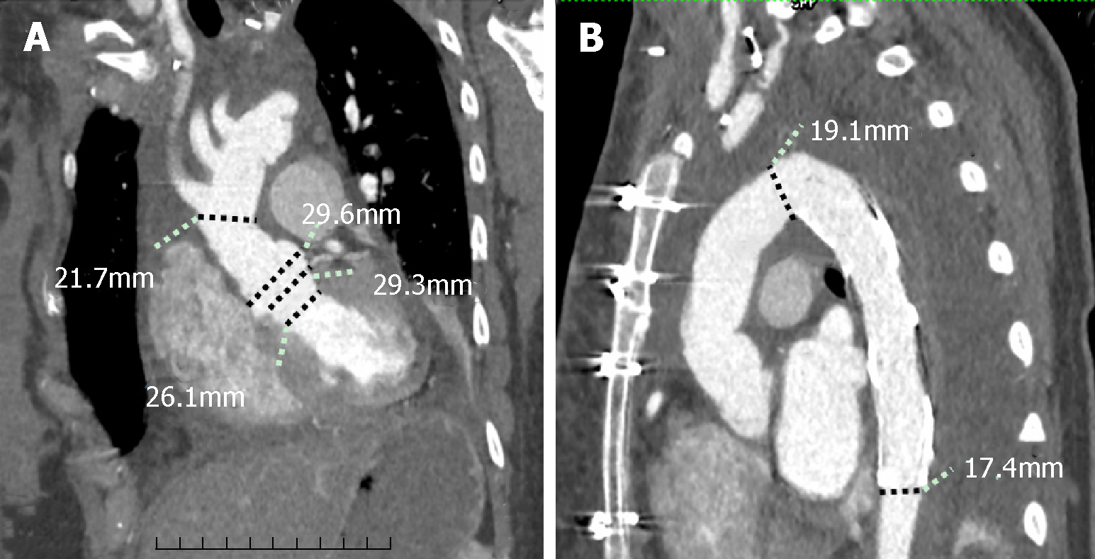
Figure 3 Postoperative images of the patient.
A and B: The good continuity of the prosthetic graft without thrombus.
- Citation: Chen SW, Zhong YL, Ge YP, Qiao ZY, Li CN, Zhu JM, Sun LZ. Successful repair of acute type A aortic dissection during pregnancy at 16th gestational week with maternal and fetal survival: A case report and review of the literature. World J Clin Cases 2019; 7(18): 2843-2850
- URL: https://www.wjgnet.com/2307-8960/full/v7/i18/2843.htm
- DOI: https://dx.doi.org/10.12998/wjcc.v7.i18.2843


