Copyright
©The Author(s) 2019.
World J Clin Cases. Sep 26, 2019; 7(18): 2802-2807
Published online Sep 26, 2019. doi: 10.12998/wjcc.v7.i18.2802
Published online Sep 26, 2019. doi: 10.12998/wjcc.v7.i18.2802
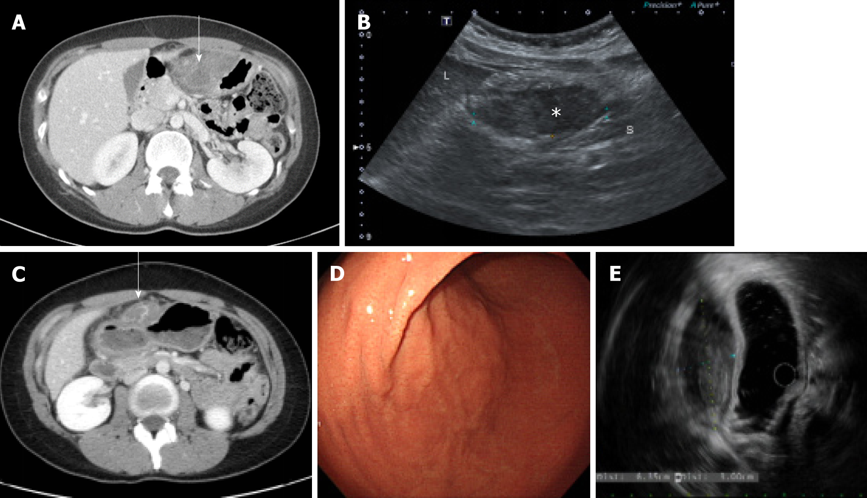
Figure 1 Imaging finding.
A: Contrast-enhanced computed tomography (CT) showed a poorly enhanced heterogenous mass adjacent to the anterior wall of the gastric lower body (arrow); B: Ultrasonography demonstrated a focally hypoechoic solid-like lesion with an internal echogenic component abutting the anterior wall of the gastric antrum (asterisk); C: Contrast-enhanced CT showed a well-circumscribed, highly attenuated, and slightly homogenous round mass on the anterior wall of the stomach (open arrow); D: Endoscopic evaluation showed a round, slightly raised area covered with normal mucosa on the anterior wall of the great curvature of the proximal antrum; E: EUS demonstrated a hypoechoic lesion of 6.35 cm × 3.00 cm.
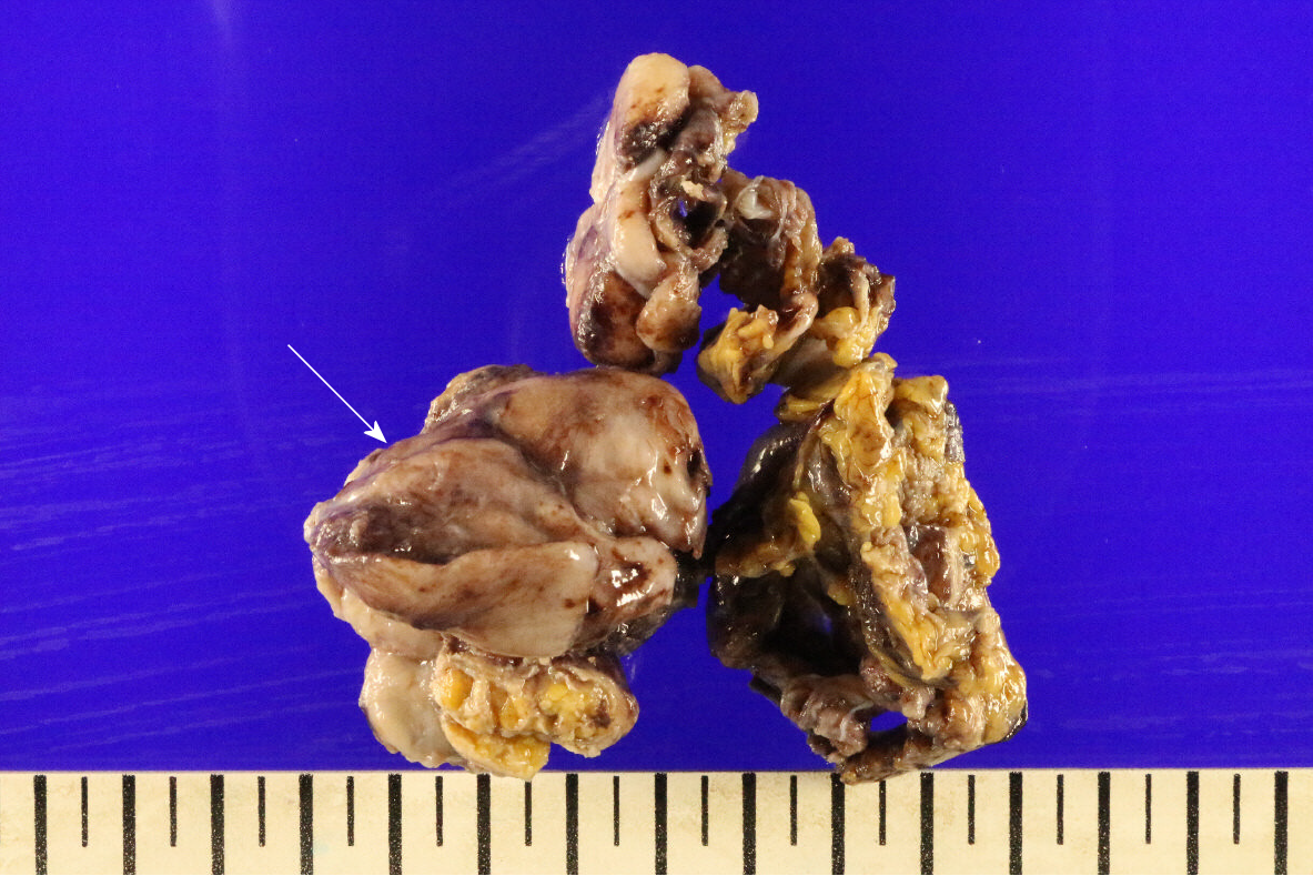
Figure 2 Gross findings of a resected specimen.
The tumor was mostly well circumscribed with a white-tan cut surface and focally infiltrative growth in fat tissue. The tumor was 6.5 cm × 4.0 cm × 1.0 cm in size (arrow).
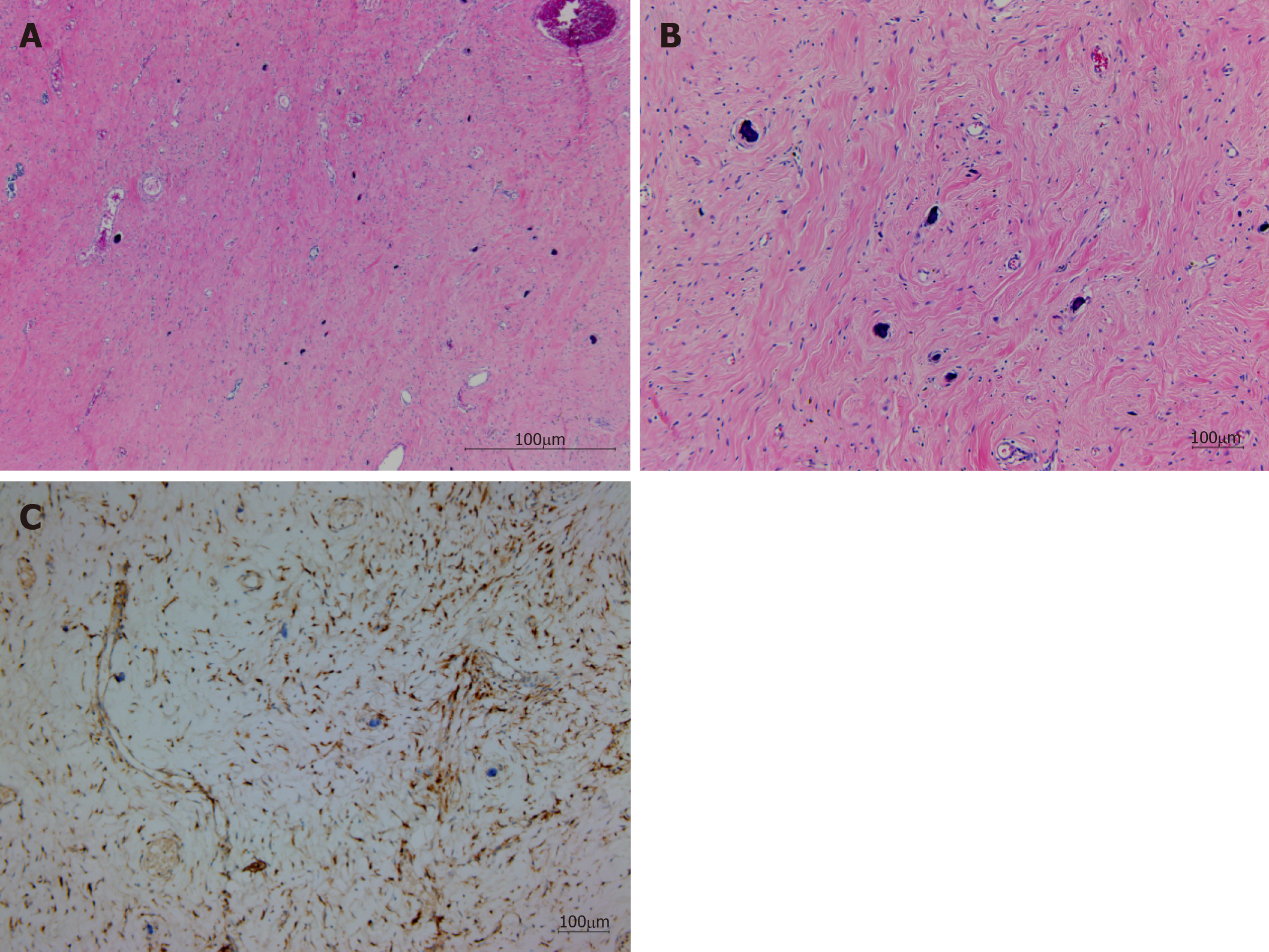
Figure 3 Histopathological result of a resected specimen.
A: Paucicellular hyalinized collagen and a haphazard collagenous matrix were observed in the tumor (H and E, × 40); B: Scattered dystrophic calcifications were observed. Lymphocytes and plasma cells were the dominant infiltrating cells, and bland spindle cells were embedded. (H and E, × 100); C: Spindle cells were positive for Factor XIIIa.
- Citation: Kwan BS, Cho DH. Calcifying fibrous tumor originating from the gastrohepatic ligament that mimicked a gastric submucosal tumor: A case report. World J Clin Cases 2019; 7(18): 2802-2807
- URL: https://www.wjgnet.com/2307-8960/full/v7/i18/2802.htm
- DOI: https://dx.doi.org/10.12998/wjcc.v7.i18.2802


