Copyright
©The Author(s) 2019.
World J Clin Cases. Sep 6, 2019; 7(17): 2463-2476
Published online Sep 6, 2019. doi: 10.12998/wjcc.v7.i17.2463
Published online Sep 6, 2019. doi: 10.12998/wjcc.v7.i17.2463
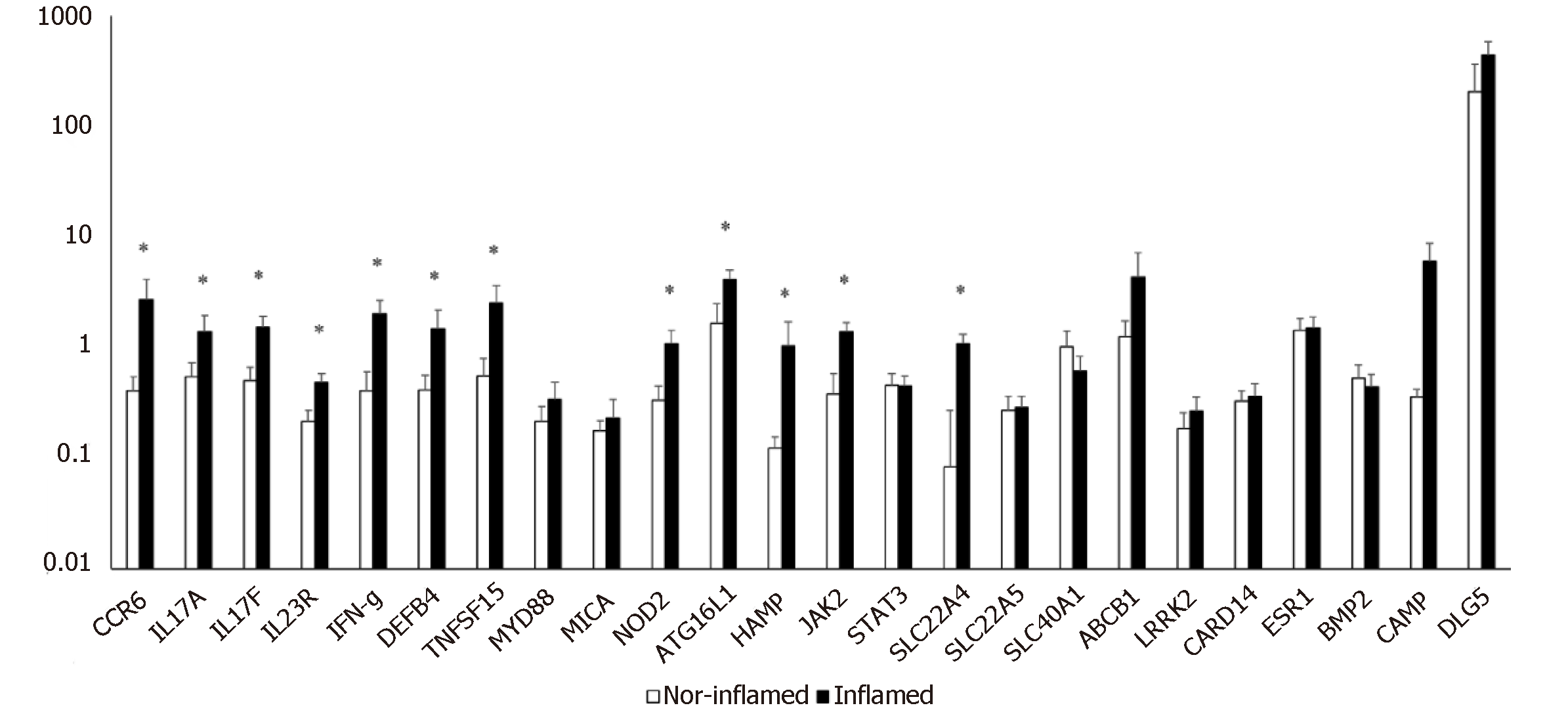
Figure 1 Quantitative evaluation of gene expression using multiplex gene assay in surgical ileum specimens of CD patients.
The abscissa shows the genes evaluated in inflamed and not inflamed tissue; the axis of the ordinates shows the value of expression of the gene normalized to the housekeeping actin (gene/β actin ratio). The first seven genes are more closely related to immunity. P value is reported only when statistically significant (P < 0.05).
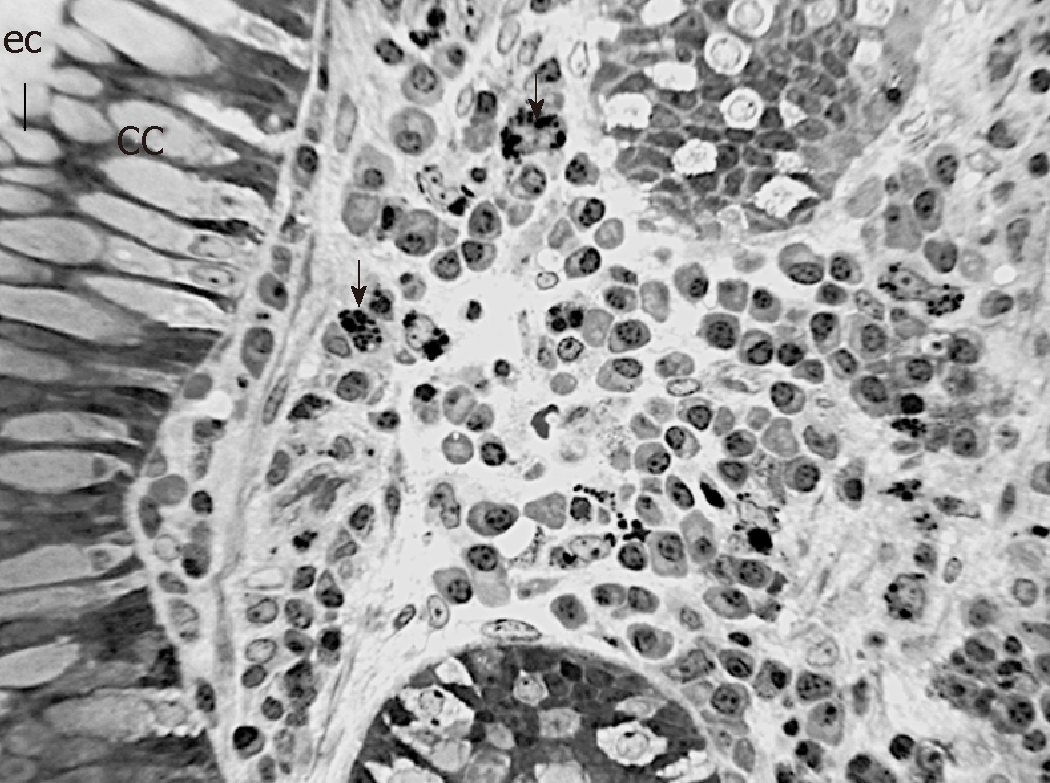
Figure 2 Representative Light microscope (LM) of the lamina propria between Lieberkühn crypts in inflamed ileal tissue of CD patient number 7b.
Notice muciparous goblet cells in cryptal epithelium and large amount and variety of immune cells (arrows) in the connective tissue. Semithin (1 µm thick) section, toluidine blue staining; cc = goblet cell, ec = enterocyte. Scale bar (4 cm) = 70 µm.
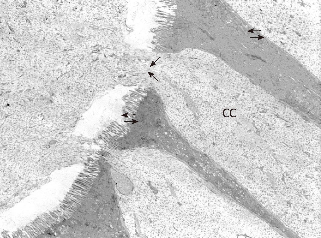
Figure 3 Representative TEM micrograph of Lieberkühn crypt wall and lumen that contains mucous product released (large arrows) by goblet cells (CC) in inflamed ileal tissue CD patient number 7b.
Small arrows indicate transport processes involving apical and lateral surfaces of enterocytes. Scale bar (1 cm) = 1 µm.
- Citation: Giudici F, Lombardelli L, Russo E, Cavalli T, Zambonin D, Logiodice F, Kullolli O, Giusti L, Bargellini T, Fazi M, Biancone L, Scaringi S, Clemente AM, Perissi E, Delfino G, Torcia MG, Ficari F, Tonelli F, Piccinni MP, Malentacchi C. Multiplex gene expression profile in inflamed mucosa of patients with Crohn’s disease ileal localization: A pilot study. World J Clin Cases 2019; 7(17): 2463-2476
- URL: https://www.wjgnet.com/2307-8960/full/v7/i17/2463.htm
- DOI: https://dx.doi.org/10.12998/wjcc.v7.i17.2463


