Copyright
©The Author(s) 2019.
World J Clin Cases. Aug 26, 2019; 7(16): 2134-2142
Published online Aug 26, 2019. doi: 10.12998/wjcc.v7.i16.2134
Published online Aug 26, 2019. doi: 10.12998/wjcc.v7.i16.2134
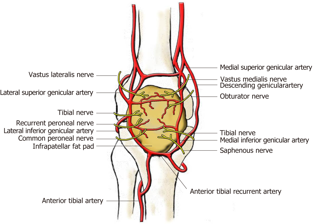
Figure 1 Blood and nerve supply of the infrapatellar fat pad (view from the front).
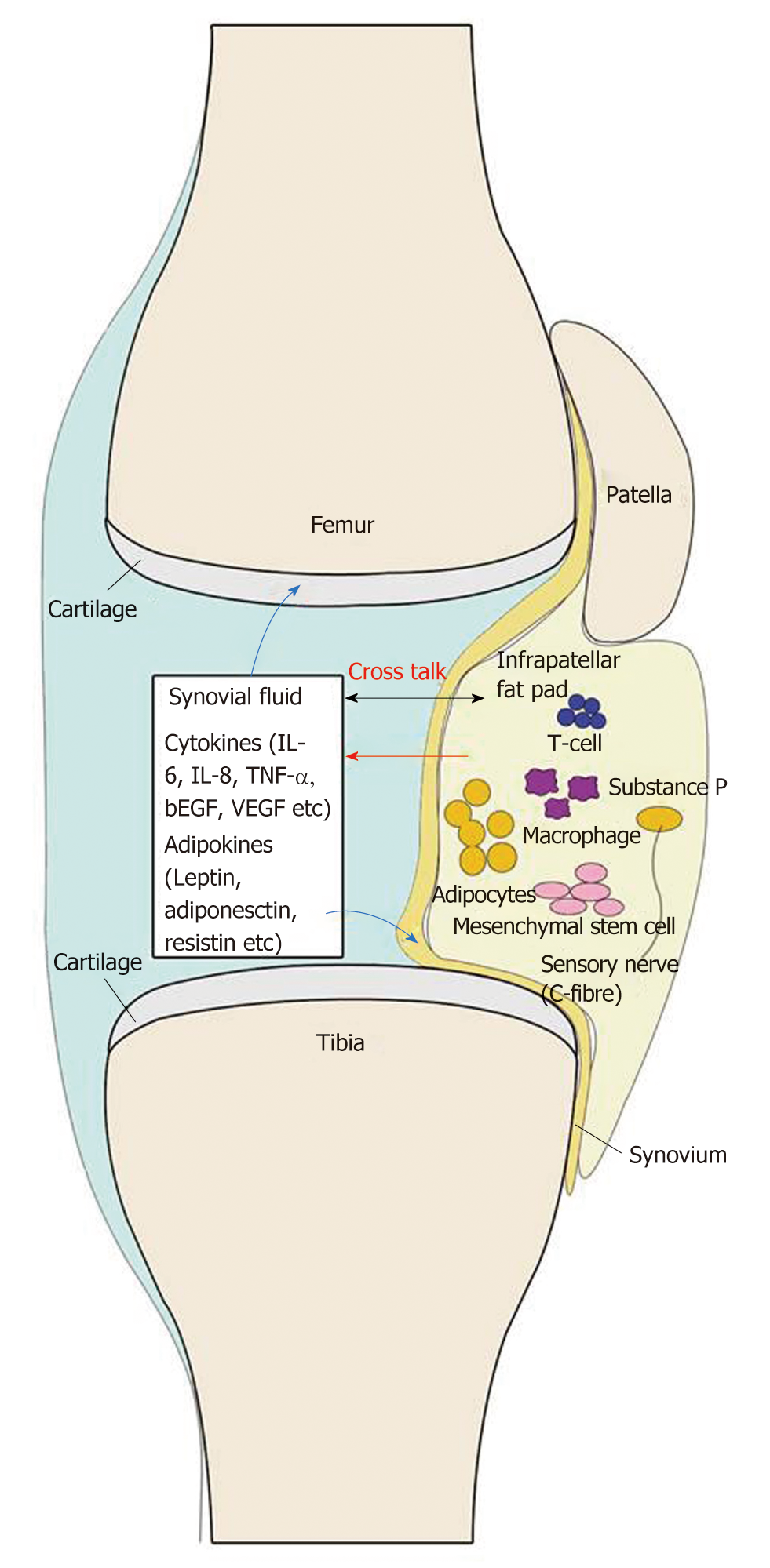
Figure 2 Current view of the infrapatellar fat pad and its interaction with other joint tissues.
The cells in the infrapatellar fat pad (IPFP) secrete cytokines, adipokines, and other factors to the synovial fluid, and then these factors react on the cells in the IPFP, synovium, and cartilage.
- Citation: Jiang LF, Fang JH, Wu LD. Role of infrapatellar fat pad in pathological process of knee osteoarthritis: Future applications in treatment. World J Clin Cases 2019; 7(16): 2134-2142
- URL: https://www.wjgnet.com/2307-8960/full/v7/i16/2134.htm
- DOI: https://dx.doi.org/10.12998/wjcc.v7.i16.2134


