Copyright
©The Author(s) 2019.
World J Clin Cases. Jul 26, 2019; 7(14): 1850-1856
Published online Jul 26, 2019. doi: 10.12998/wjcc.v7.i14.1850
Published online Jul 26, 2019. doi: 10.12998/wjcc.v7.i14.1850
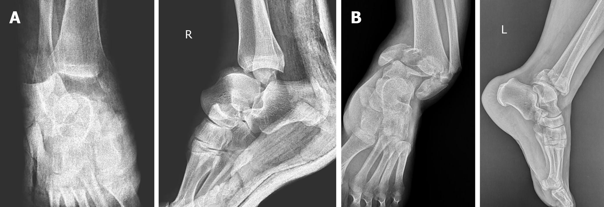
Figure 1 Anteroposterior and lateral ankle radiographs of the right ankle (A) showing anterolateral total talus dislocation accompanied by a fracture of the medial malleolus and pilon fracture of the left ankle (B).
Figure 2 Computed tomography images after unsuccessful closed reduction showing fractured highly unstable medial malleolus displaced into the ankle mortise, blocking the relocation of the talus.
A: Coronal view demonstrating the extent of extrusion of the talus under the tibial plafond; B: Sagittal view demonstrating the medial malleolus in the ankle mortise (arrow); C and D: Coronal and axial views demonstrating the displaced medial malleolus blocking the talus (arrows).
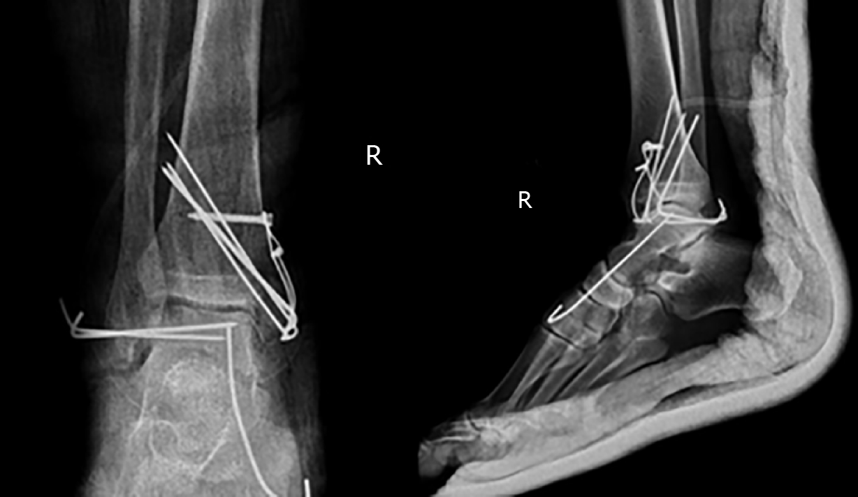
Figure 3 Anteroposterior and lateral ankle radiographs of the right ankle after surgery.
After the reduction of the medial malleolus and the talus, the medial malleolus was fixed using the tension band technique because of instability of the tibiotalar and talonavicular joints. Two Kirschner (K) wires were inserted horizontally from the lateral malleolus to the talus in order to increase the stability of the tibiotalar joint and a K-wire for the talonavicular joint was used for the maintenance of the reduction.
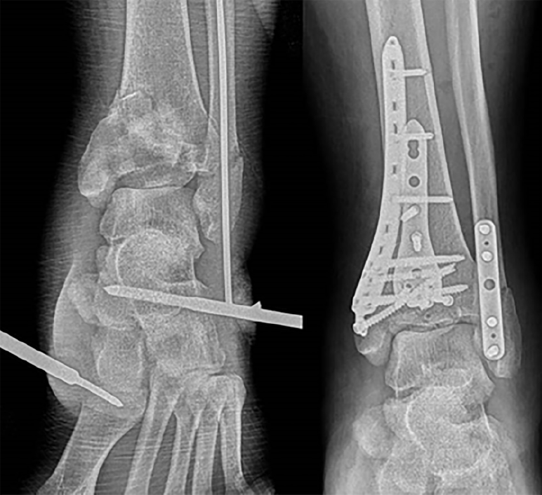
Figure 4 Anteroposterior ankle radiographs of the left ankle after surgery on the left and after two weeks on the right.
Due to the swelling in the left ankle, external fixation was performed in order to restore the alignment and open reduction, and internal fixation with locking anatomical plates and screws using anterolateral and posterolateral approaches was performed two weeks after the initial surgery.
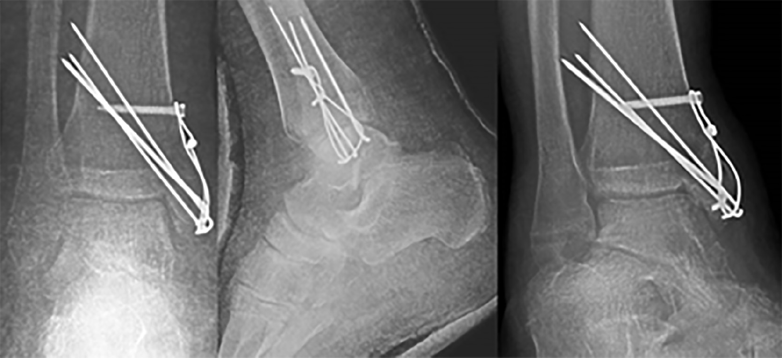
Figure 5 AP, mortise, and lateral ankle radiographs of the right ankle after removal of the talofibular and talonavicular K-wires at the 6th postoperative week.
Note that there was no Hawkins sign.
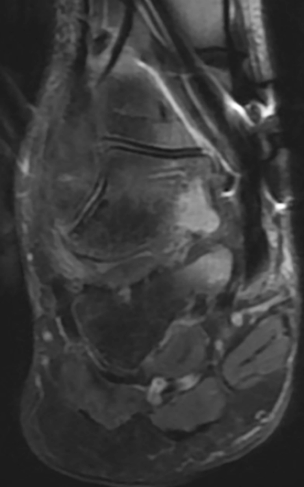
Figure 6 Magnetic resonance imaging at 6 weeks revealing only talar bone bruising and edema but no signs of avascular necrosis.
- Citation: Yapici F, Coskun M, Arslan MC, Ulu E, Akman YE. Open reduction of a total talar dislocation: A case report and review of the literature. World J Clin Cases 2019; 7(14): 1850-1856
- URL: https://www.wjgnet.com/2307-8960/full/v7/i14/1850.htm
- DOI: https://dx.doi.org/10.12998/wjcc.v7.i14.1850


