Copyright
©The Author(s) 2019.
World J Clin Cases. Jul 26, 2019; 7(14): 1837-1843
Published online Jul 26, 2019. doi: 10.12998/wjcc.v7.i14.1837
Published online Jul 26, 2019. doi: 10.12998/wjcc.v7.i14.1837
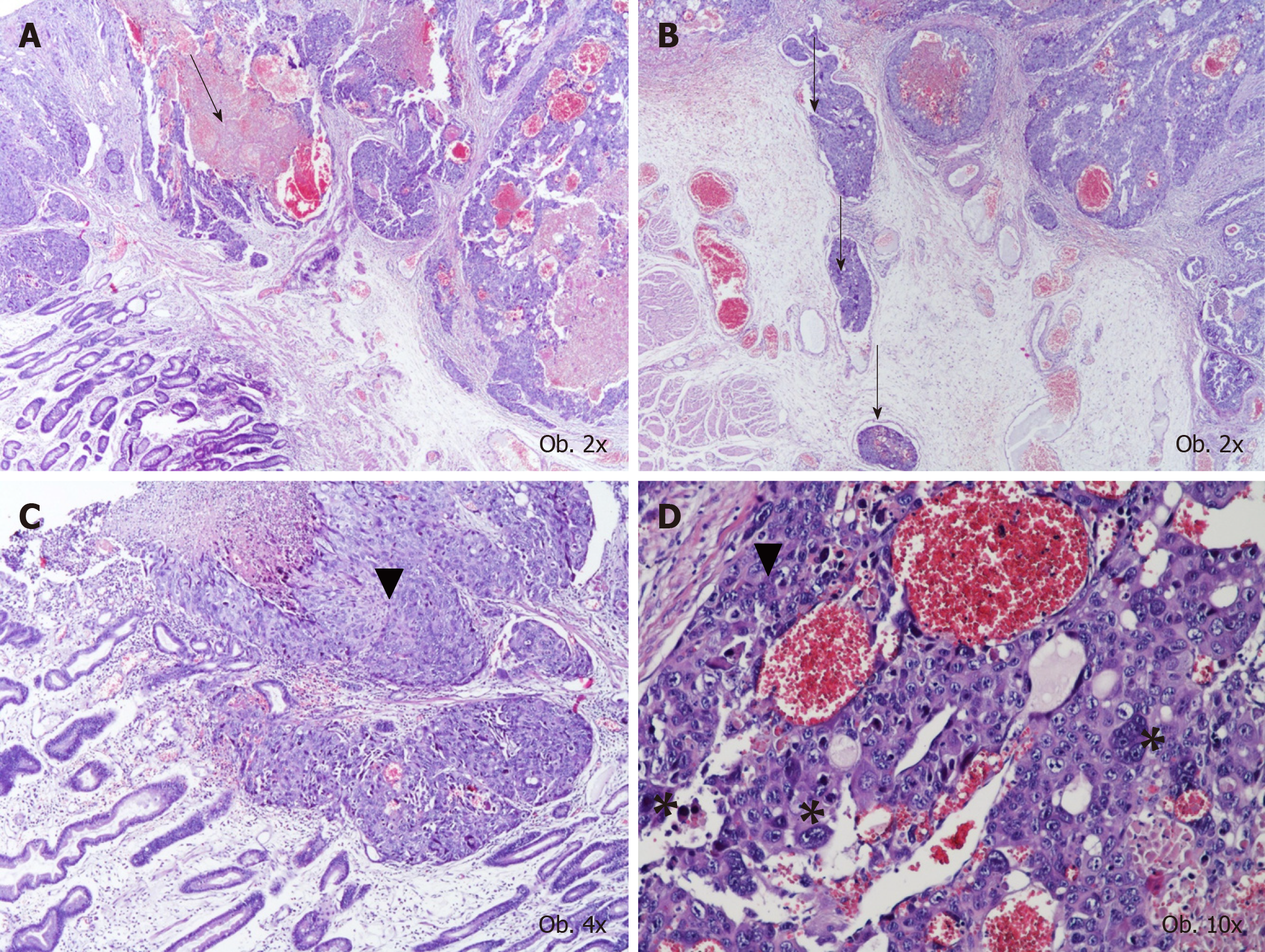
Figure 1 Histological examination results.
A, B: The histological aspect of choriocarcinoma, with large hemorrhagic area (A), multiple vascular emboli (B), and characteristic proliferation of oval cells with pale cytoplasm with similar aspect to cytotrophoblastic cells (marked by ▼) and giant cells with lobulated or bizarre nuclei (marked by *); C, D: Similar to syncytiotrophoblastic cells.
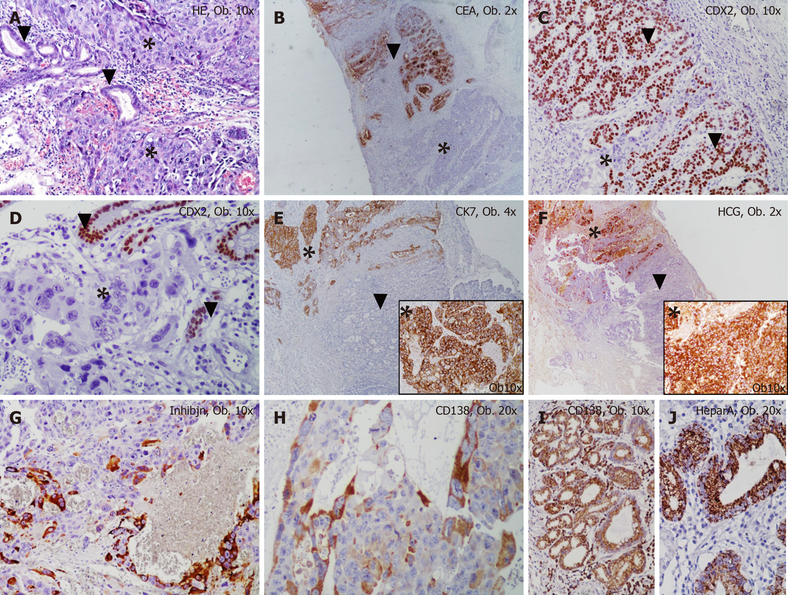
Figure 2 Histological examination results.
A: The mixed gastric carcinoma shows two components: choriocarcinoma (marked by *) and adenocarcinoma (marked by ▼); B-F: The choriocarcinoma cells are negative for CEA (B) and CDX2 (C, D) and show positivity for Cytokeratin 7 (E) and HCG (F); G, H: The syncytiotrophoblast-like cells are marked by inhibin (G) and CD138 (H). The adenocarcinoma component is positive for CEA (B) and CDX2 (C, D) and to not present positivity for Cytokeratin 7 (E) and HCG (F); I, J: The normal gastric mucosa cells are marked by CD138 (I) and Hepar A (J).
- Citation: Gurzu S, Copotoiu C, Tugui A, Kwizera C, Szodorai R, Jung I. Primary gastric choriocarcinoma - a rare and aggressive tumor with multilineage differentiation: A case report. World J Clin Cases 2019; 7(14): 1837-1843
- URL: https://www.wjgnet.com/2307-8960/full/v7/i14/1837.htm
- DOI: https://dx.doi.org/10.12998/wjcc.v7.i14.1837


