Copyright
©The Author(s) 2019.
World J Clin Cases. Jul 6, 2019; 7(13): 1652-1659
Published online Jul 6, 2019. doi: 10.12998/wjcc.v7.i13.1652
Published online Jul 6, 2019. doi: 10.12998/wjcc.v7.i13.1652
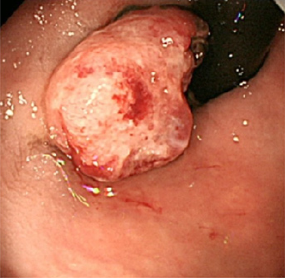
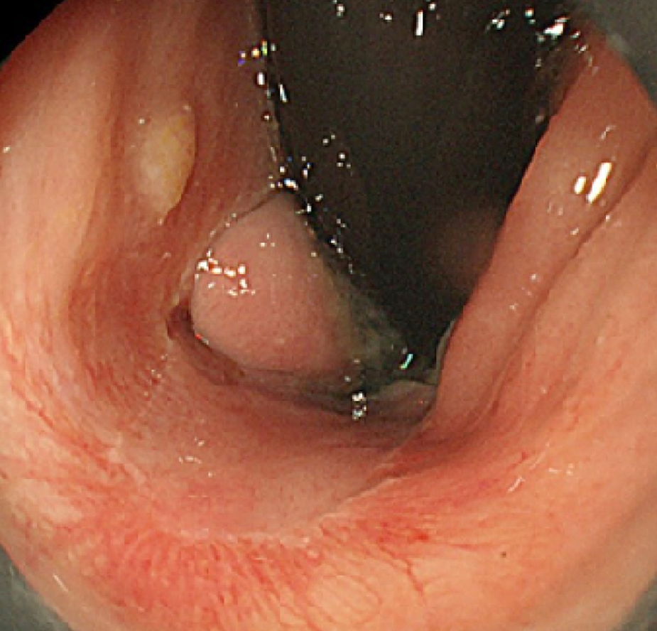
Figure 1 Colonoscopic findings.
A 20-mm protruded lesion in the lower rectum just above the dentate line is detected.
Figure 2 Narrow-band imaging with magnifying endoscopy findings.
Non-structural areas and some thick, unevenly sized vessels with branching and curtailed irregularity are observed.
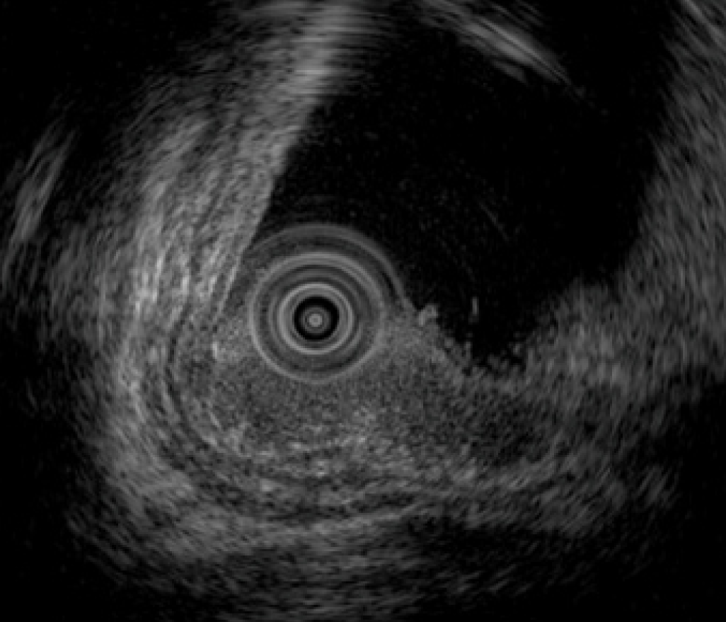
Figure 3 Endoscopic ultrasonography findings.
Endoscopic ultrasonography image shows the lesion as a hypoechoic mass. The tumor invades the submucosal layer but not the muscular layer.
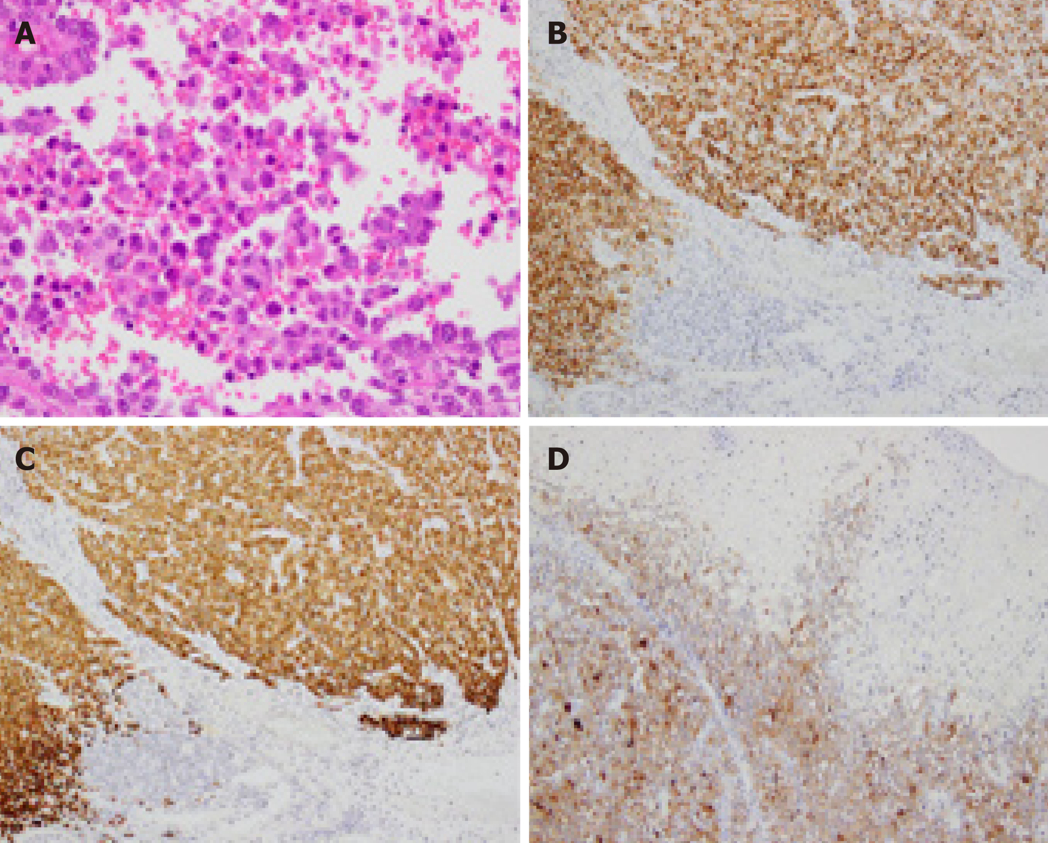
Figure 4 Histopathological and immunohistochemical findings of the endoscopic submucosal dissection-resected specimen.
A: Hematoxylin and eosin staining shows diffuse infiltration of round tumor cells with irregular nuclei. Melanin pigment is absent (magnification × 400). The tumor cells are positive for B: Human Melanin Black-45 (× 200), C: Melan-A (× 200), and D: S-100 (× 100).
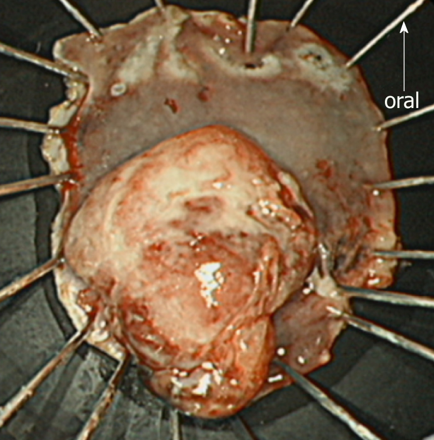
Figure 5 Endoscopic submucosal dissection-resected specimen.
The endoscopic submucosal dissection-resected specimen is 40 × 30 mm in size. The tumor is protruded and measures 20 × 15 mm. Macroscopically, an adequate margin from the tumor, especially on the oral side, is ensured.
Figure 6 Follow-up endoscopic findings 3 mo after the endoscopic submucosal dissection.
The post-endoscopic submucosal dissection ulcer is healed, and no local recurrence is detected.
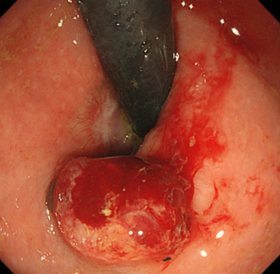
Figure 7 Follow-up endoscopic findings 6 mo after the endoscopic submucosal dissection.
A 10-mm protruded lesion is identified about 10 mm from the oral side of the post-endoscopic submucosal dissection scar. Biopsy specimens from this lesion confirm a malignant melanoma.
- Citation: Manabe S, Boku Y, Takeda M, Usui F, Hirata I, Takahashi S. Endoscopic submucosal dissection as excisional biopsy for anorectal malignant melanoma: A case report. World J Clin Cases 2019; 7(13): 1652-1659
- URL: https://www.wjgnet.com/2307-8960/full/v7/i13/1652.htm
- DOI: https://dx.doi.org/10.12998/wjcc.v7.i13.1652


