Copyright
©The Author(s) 2019.
World J Clin Cases. Jun 6, 2019; 7(11): 1323-1329
Published online Jun 6, 2019. doi: 10.12998/wjcc.v7.i11.1323
Published online Jun 6, 2019. doi: 10.12998/wjcc.v7.i11.1323
Figure 1 Multi-hole self-expandable metallic stent.
A: With lasso (small-hole type); B: Large-hole type.
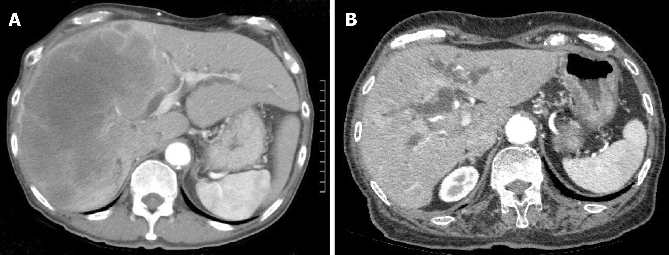
Figure 2 Computerized tomography images.
A: A large metastasis in the right liver lobe; B: Intrahepatic biliary dilation.
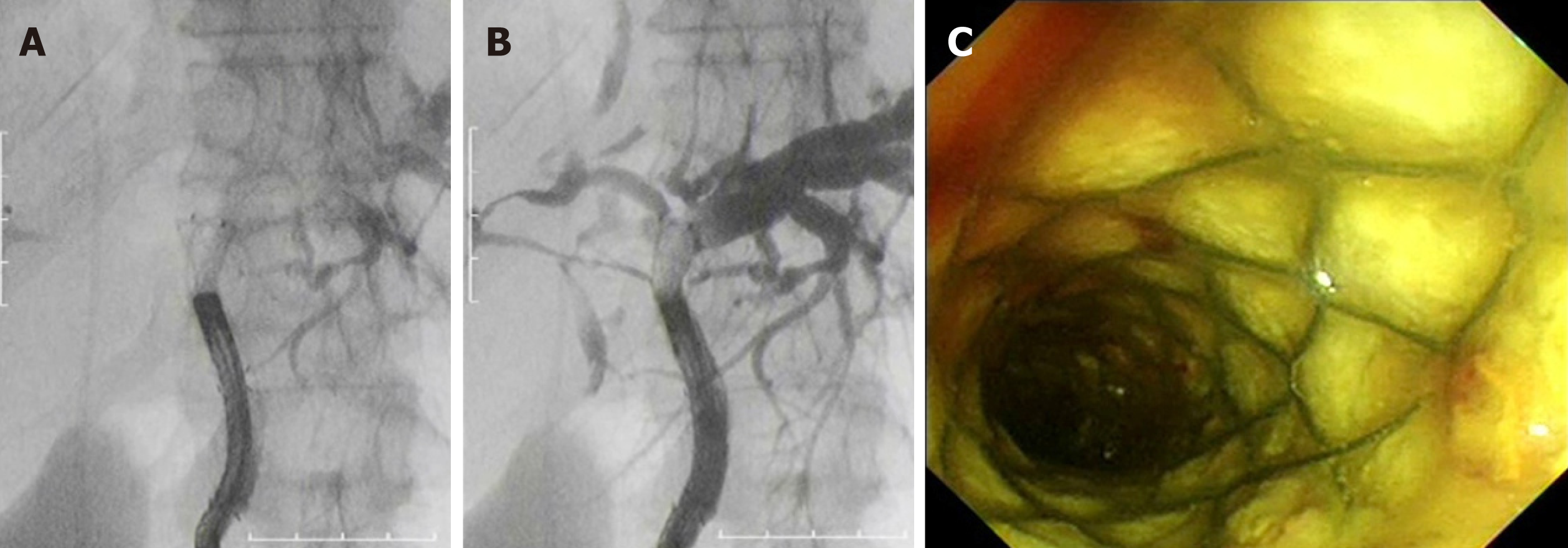
Figure 3 Cholangiography via a direct peroral cholangioscope.
A, B: The right bile duct was visualized by contrast material through the stent openings; C: After cholangiography, the metallic mesh was not found buried in tissue but fixed to the bile duct wall.
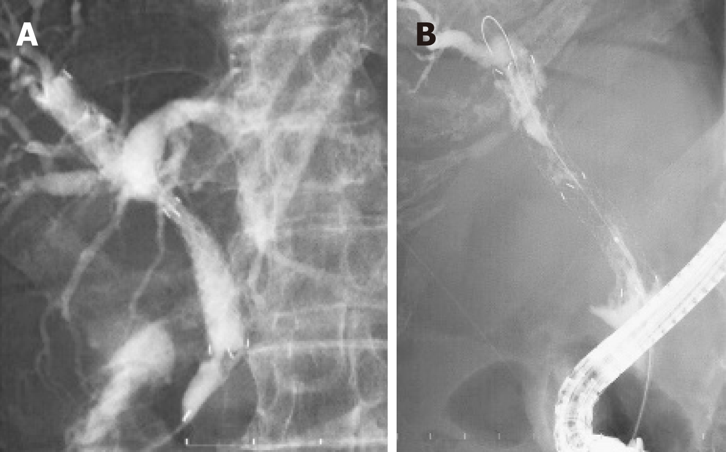
Figure 4 Cholangiography of a multi-hole self-expandable metallic stent and ablation therapy.
A: The left bile duct was visualized by contrast material through the stent openings; B: Ablation therapy by monopolar catheter and endoscopic retrograde cholangiography.
- Citation: Kobayashi M. Development of a biliary multi-hole self-expandable metallic stent for bile tract diseases: A case report. World J Clin Cases 2019; 7(11): 1323-1329
- URL: https://www.wjgnet.com/2307-8960/full/v7/i11/1323.htm
- DOI: https://dx.doi.org/10.12998/wjcc.v7.i11.1323


