Copyright
©The Author(s) 2019.
World J Clin Cases. May 26, 2019; 7(10): 1213-1220
Published online May 26, 2019. doi: 10.12998/wjcc.v7.i10.1213
Published online May 26, 2019. doi: 10.12998/wjcc.v7.i10.1213
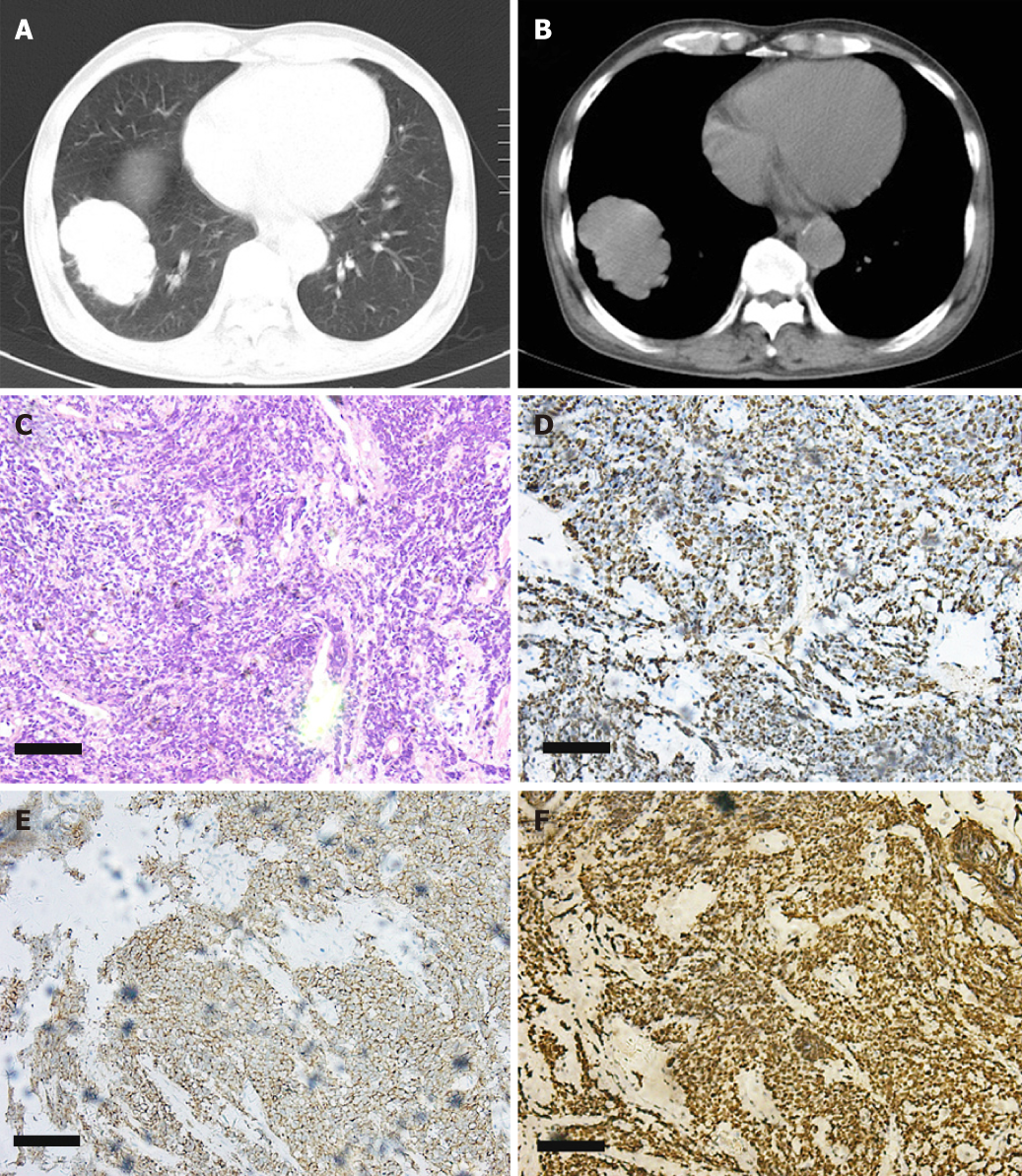
Figure 1 Lung computed tomography and bronchoscopic biopsy of Case 1.
A, B: Lung computed tomography of the patient. Right middle lobe: Peripheral lung cancer with lymph node metastasis and distal obstructive pneumonia. Bilateral pleural effusion; C: Hematoxylin and eosin staining of the tissue; D: Ki-67 staining of the tissue; E: Synaptophysin staining of the tissue; F: Thyroid transcription factor-1 staining of the tissue.
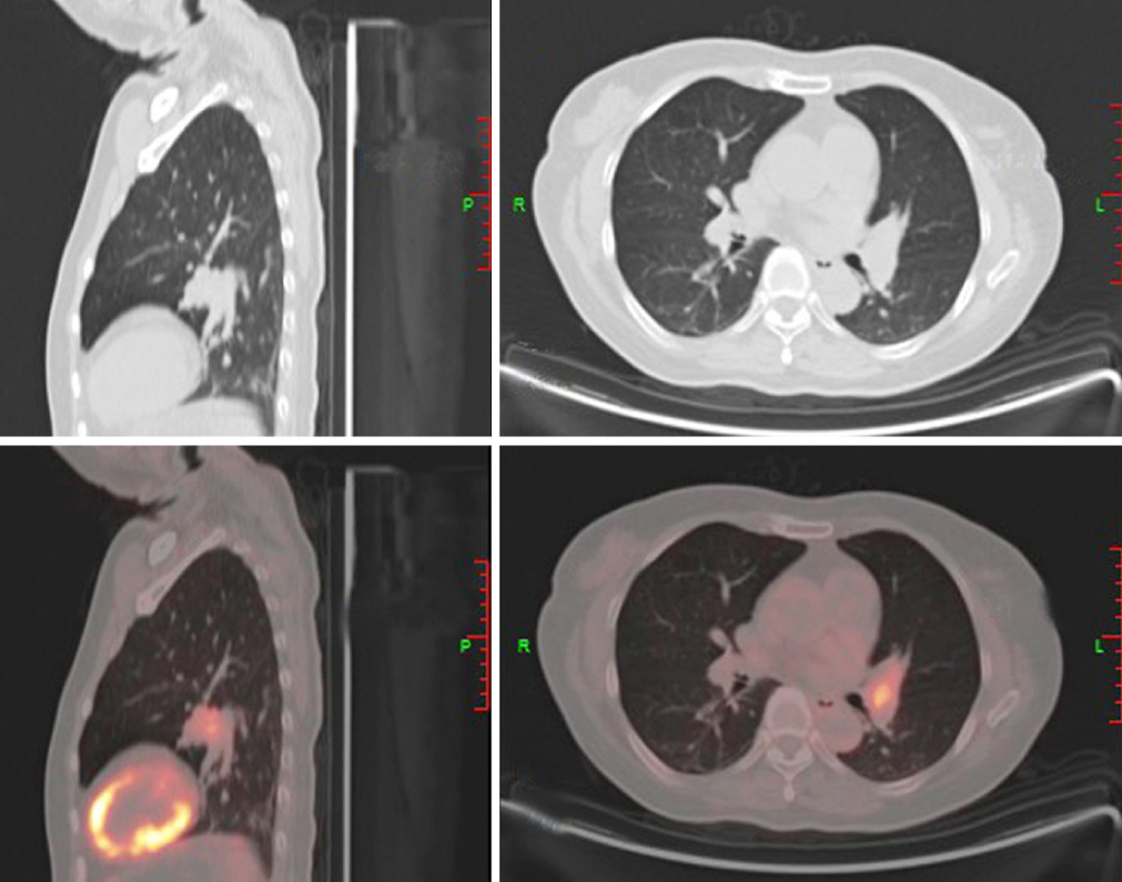
Figure 2 Positron emission tomography-computed tomography of Case 2.
A hypermetabolic nodule is visible in the left lingular lobe (central lung cancer).
- Citation: Zhou T, Wang Y, Zhao X, Liu Y, Wang YX, Gang XK, Wang GX. Small cell lung cancer starting with diabetes mellitus: Two case reports and literature review. World J Clin Cases 2019; 7(10): 1213-1220
- URL: https://www.wjgnet.com/2307-8960/full/v7/i10/1213.htm
- DOI: https://dx.doi.org/10.12998/wjcc.v7.i10.1213


