Copyright
©The Author(s) 2019.
World J Clin Cases. May 26, 2019; 7(10): 1184-1190
Published online May 26, 2019. doi: 10.12998/wjcc.v7.i10.1184
Published online May 26, 2019. doi: 10.12998/wjcc.v7.i10.1184
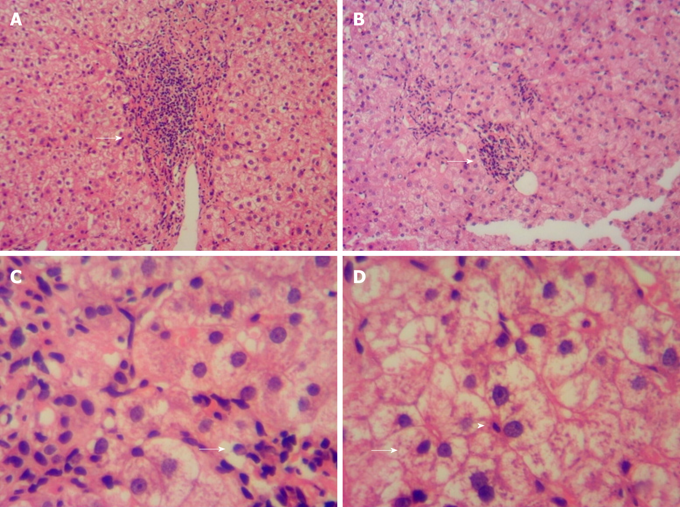
Figure 1 Liver histology (hematoxylin and eosin staining).
A: Mild interfacial inflammation and in portal area; B: Focal necrosis (arrows, 10 × 20); C: lymphatic-plasma cell infiltration and plasma cell infiltration (arrows, 10 × 20); and D: Rosetting of hepatocytes (arrows) and lymphocyte penetration (arrowheads, 10 × 40).
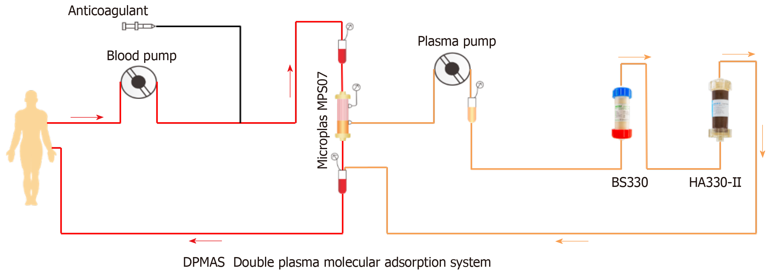
Figure 2 Double plasma molecular absorption system flowchart.
- Citation: Tan YW, Sun L, Zhang K, Zhu L. Therapeutic plasma exchange and a double plasma molecular absorption system in the treatment of thyroid storm with severe liver injury: A case report. World J Clin Cases 2019; 7(10): 1184-1190
- URL: https://www.wjgnet.com/2307-8960/full/v7/i10/1184.htm
- DOI: https://dx.doi.org/10.12998/wjcc.v7.i10.1184


