Copyright
©The Author(s) 2019.
World J Clin Cases. May 26, 2019; 7(10): 1142-1148
Published online May 26, 2019. doi: 10.12998/wjcc.v7.i10.1142
Published online May 26, 2019. doi: 10.12998/wjcc.v7.i10.1142
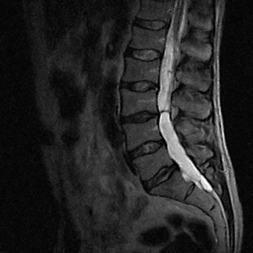
Figure 1 The first magnetic resonance imaging of the lumbosacral spine in 2007 showing only slight degenerative changes.
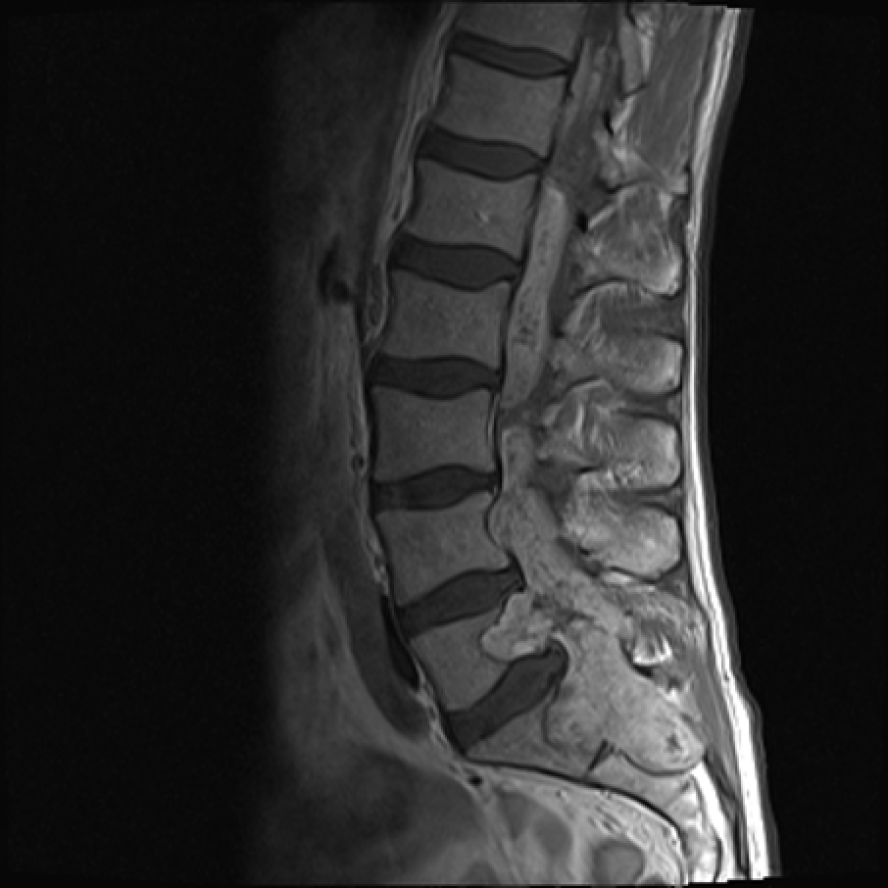
Figure 2 An extensive tumorous lesion was seen in 2014, located intramedullary, extending from the Th11, invading the conus medullaris and encompassing the entire sacral and lumbar canal to the S2 level, invading the vertebrae and spreading to perispinal muscles.
Homogenous contrast enhancement is evident.
Figure 3 Histological examination results.
A: Myxopapillary ependymomas display a variable papillary architecture with cuboid to elongated glial cells radially arranged in myxoid stroma with a central blood vessel; B: The tumour cells are immunoreactive for vimentin and GFAP; C: Immunoreactivity for cytokeratins is typically absent.
Figure 4 The control magnetic resonance imaging showing no progression of the tumour.
- Citation: Strojnik T, Bujas T, Velnar T. Invasive myxopapillary ependymoma of the lumbar spine: A case report. World J Clin Cases 2019; 7(10): 1142-1148
- URL: https://www.wjgnet.com/2307-8960/full/v7/i10/1142.htm
- DOI: https://dx.doi.org/10.12998/wjcc.v7.i10.1142


