Copyright
©The Author(s) 2019.
World J Clin Cases. Jan 6, 2019; 7(1): 89-94
Published online Jan 6, 2019. doi: 10.12998/wjcc.v7.i1.89
Published online Jan 6, 2019. doi: 10.12998/wjcc.v7.i1.89
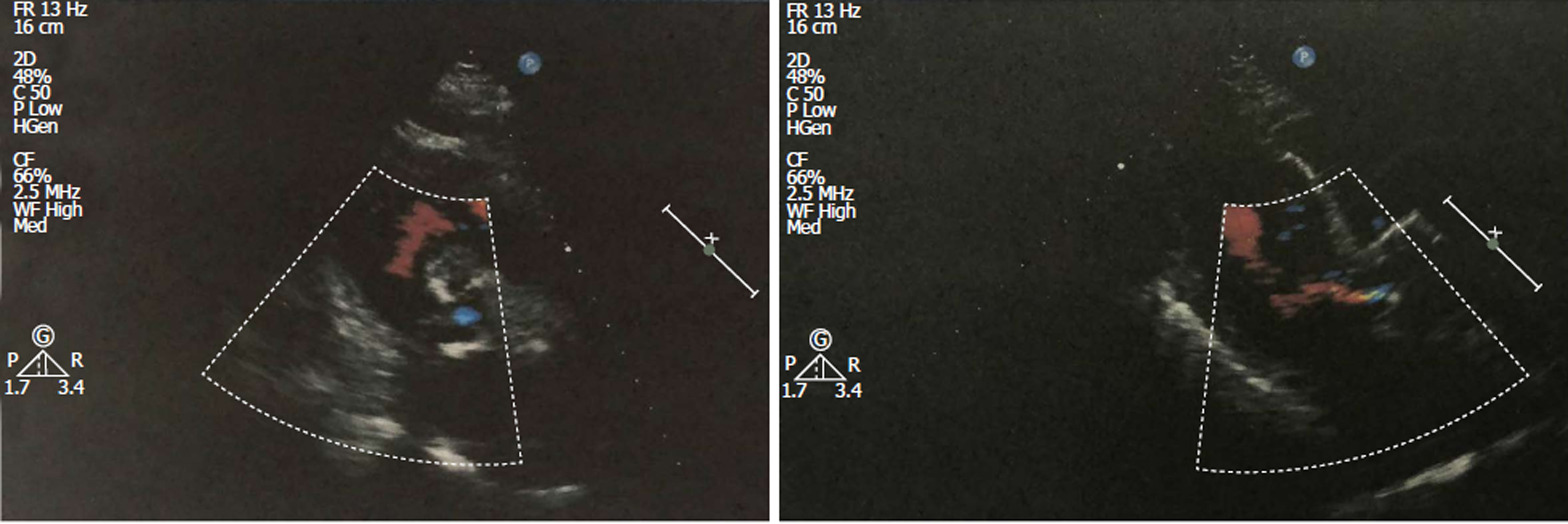
Figure 1 Echocardiographic examination on October 28, 2017.
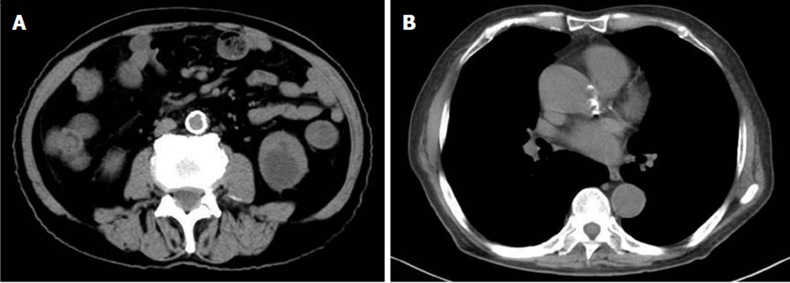
Figure 2 Pre-surgery computed tomography examination.
A: A soft tissue shadow of the ileocecal junction and adjacent ileum surrounded by multiple swollen lymph nodes; B: Aortic and coronary atherosclerosis.
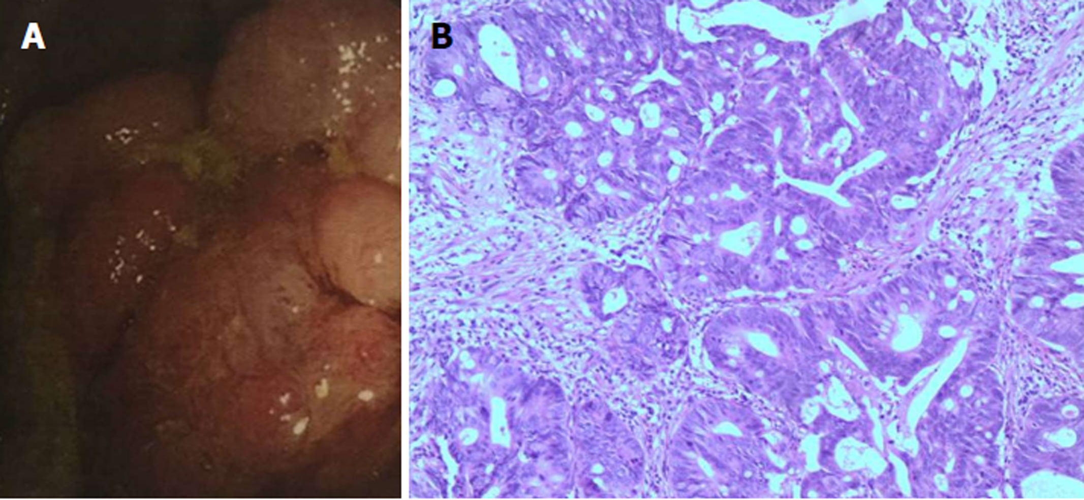
Figure 3 Diagnosis of a space-occupying ileocecal lesion.
A: Colonoscopic examination; B: H&E staining showed adenocarcinoma (magnification, × 100).
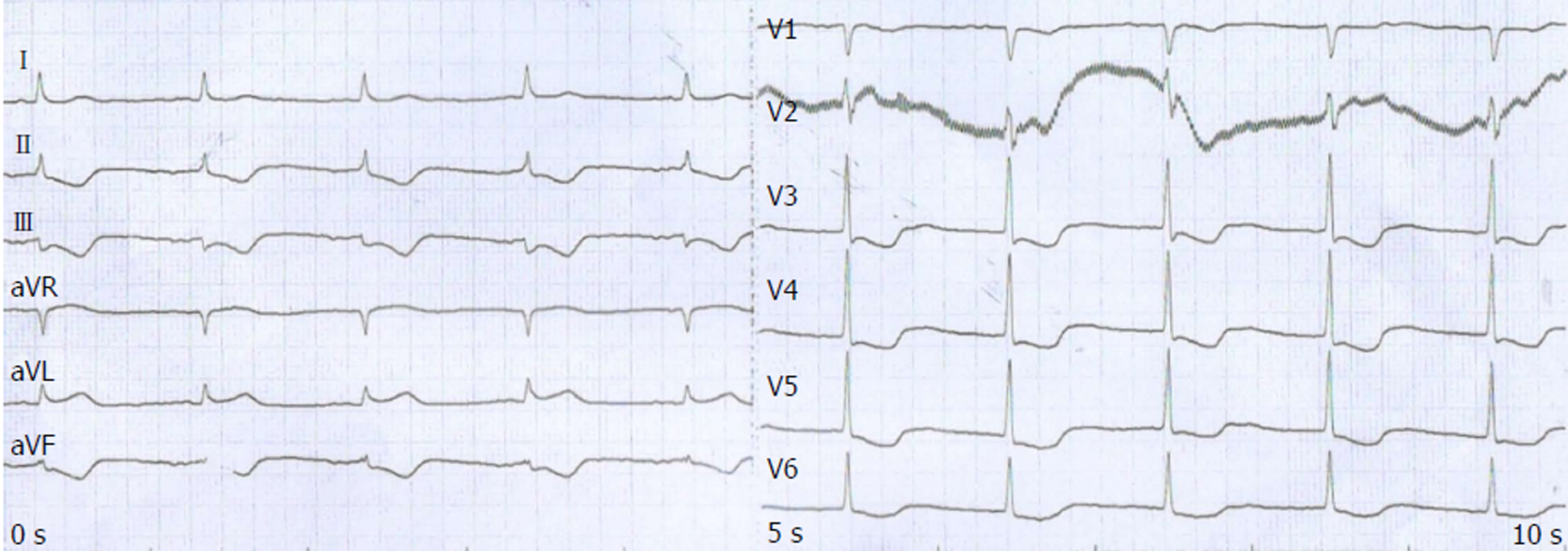
Figure 4 Electrocardiogram results on October 28, 2017.
- Citation: Zhang SC, Yu MY, Xi L, Zhang JX. Tegafur deteriorates established cardiovascular atherosclerosis in colon cancer: A case report and review of the literature. World J Clin Cases 2019; 7(1): 89-94
- URL: https://www.wjgnet.com/2307-8960/full/v7/i1/89.htm
- DOI: https://dx.doi.org/10.12998/wjcc.v7.i1.89


