Copyright
©The Author(s) 2019.
World J Clin Cases. Jan 6, 2019; 7(1): 102-108
Published online Jan 6, 2019. doi: 10.12998/wjcc.v7.i1.102
Published online Jan 6, 2019. doi: 10.12998/wjcc.v7.i1.102
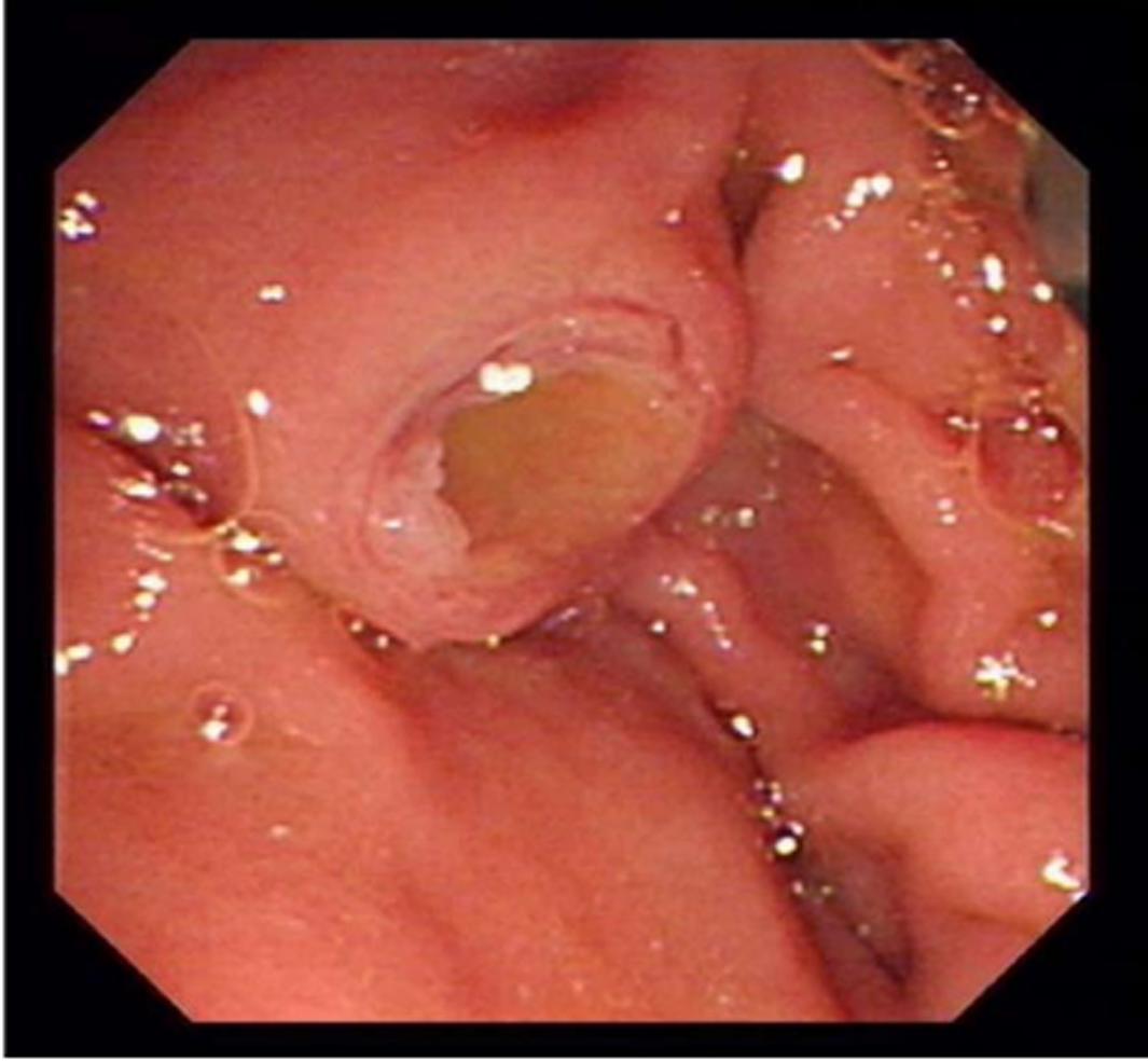
Figure 1 Image acquired during endoscopic retrograde cholangiopancreatography.
Duodenoscopy revealed duodenal major papilla with fish eye-like appearance.
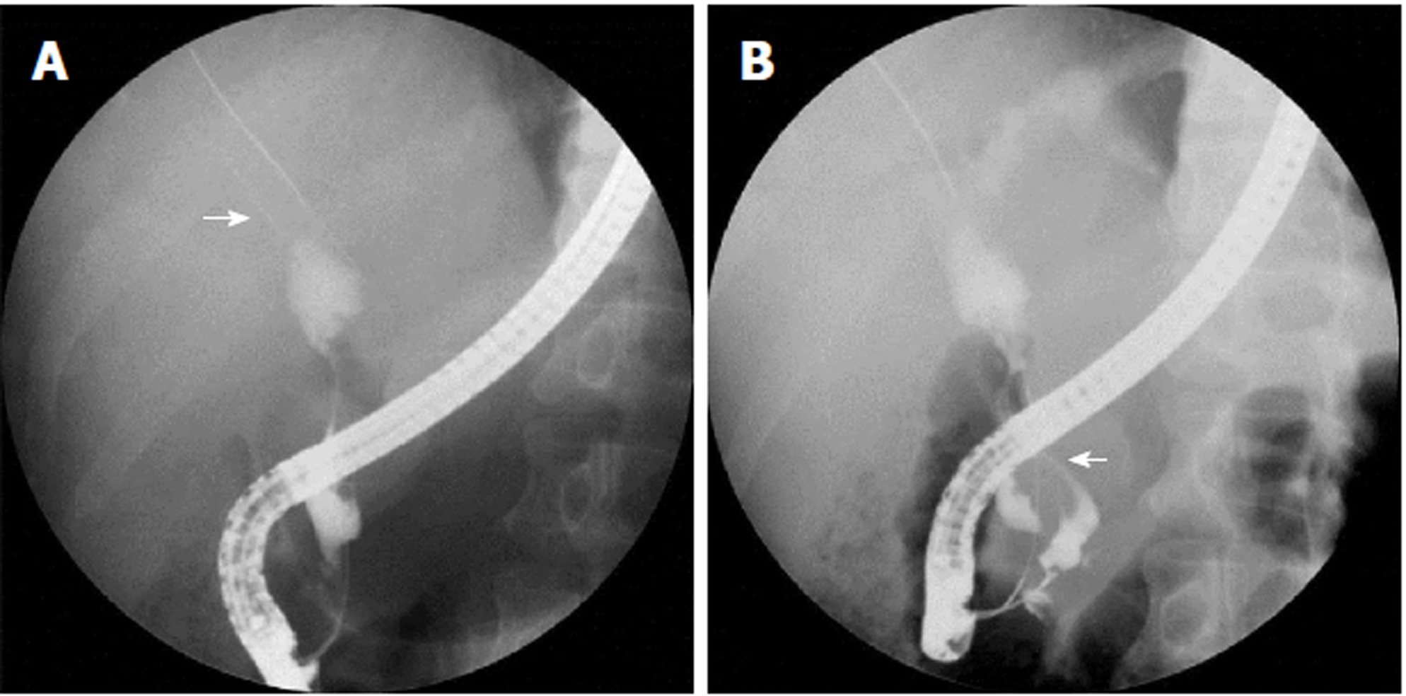
Figure 2 Endoscopic retrograde cholangiopancreatography images.
A: Cholangiogram showed common bile duct stricture with proximal bile duct dilation and a metal stent that displaced into the intrahepatic bile duct (indicated by arrows); B: Pancreatography revealed a pancreatobiliary fistula connected to the pancreatic and bile duct (indicated by arrows).
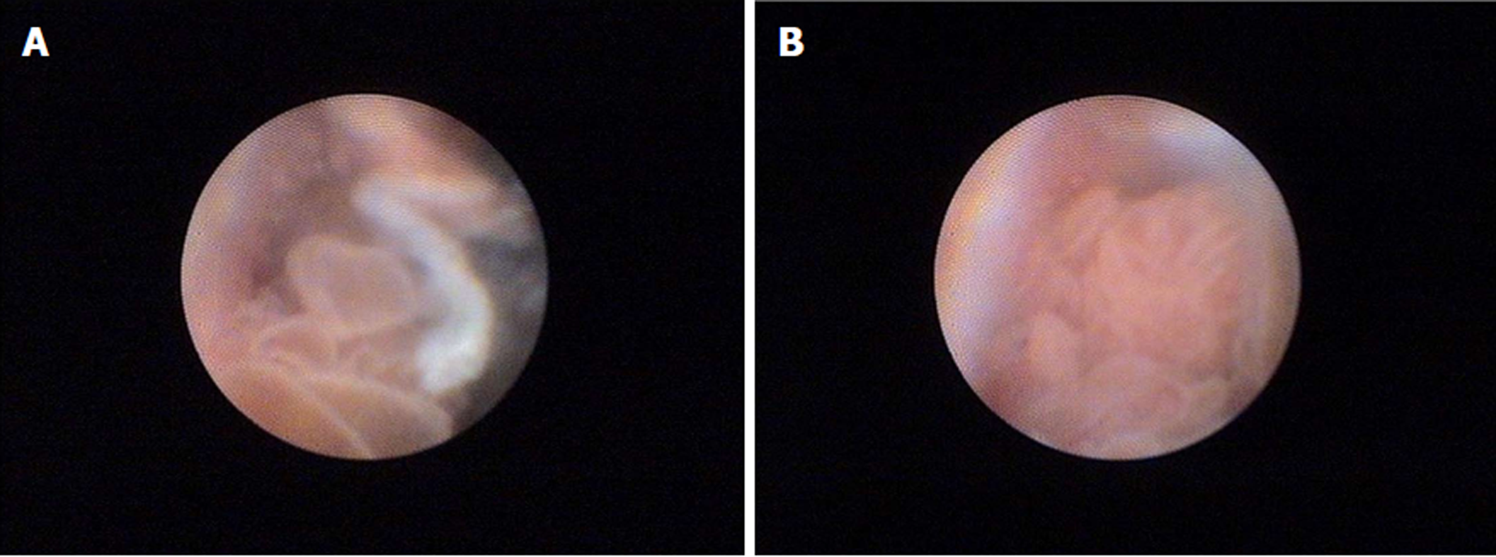
Figure 3 SpyGlass images.
A: Multiple fish egg-like protrusions in the common bile duct; B: A cauliflower-like protrusion with vascular images in the common bile duct.
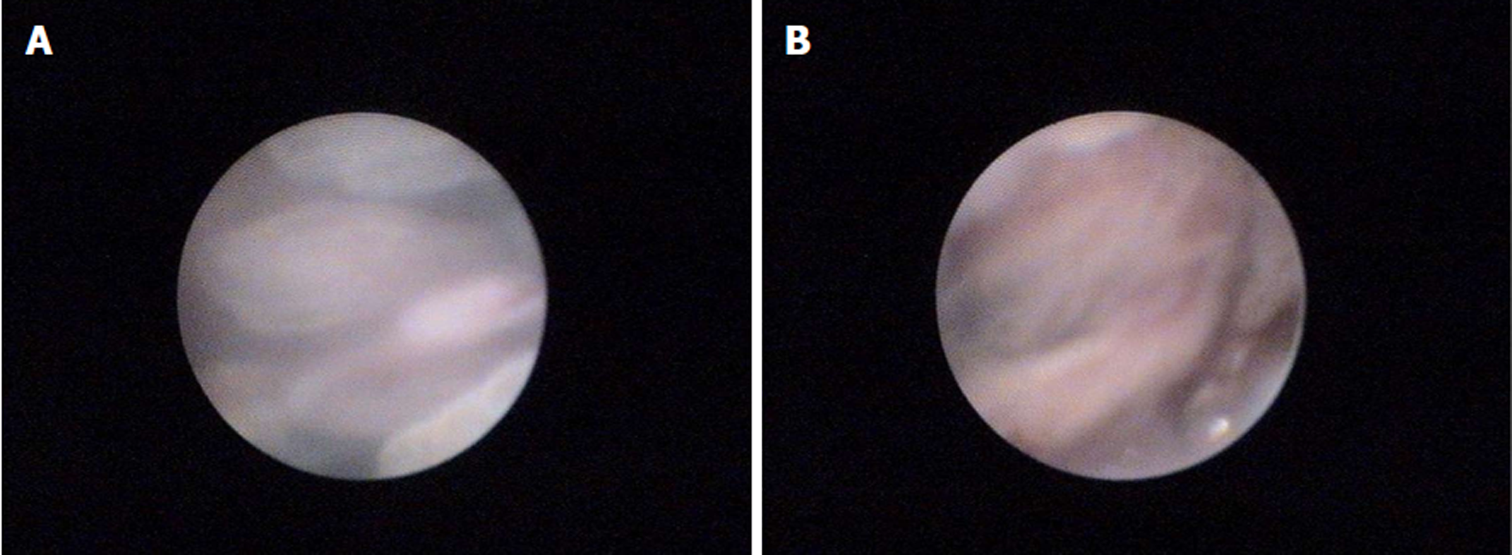
Figure 4 SpyGlass images.
A: Multiple high papillary protrusions in the main pancreatic duct; B: Leaf-like projections with vascular images in the main pancreatic duct.
- Citation: Ren X, Zhu CL, Qin XF, Jiang H, Xia T, Qu YP. Co-occurrence of IPMN and malignant IPNB complicated by a pancreatobiliary fistula: A case report and review of the literature. World J Clin Cases 2019; 7(1): 102-108
- URL: https://www.wjgnet.com/2307-8960/full/v7/i1/102.htm
- DOI: https://dx.doi.org/10.12998/wjcc.v7.i1.102


