Copyright
©The Author(s) 2019.
World J Clin Cases. Jan 6, 2019; 7(1): 10-18
Published online Jan 6, 2019. doi: 10.12998/wjcc.v7.i1.10
Published online Jan 6, 2019. doi: 10.12998/wjcc.v7.i1.10
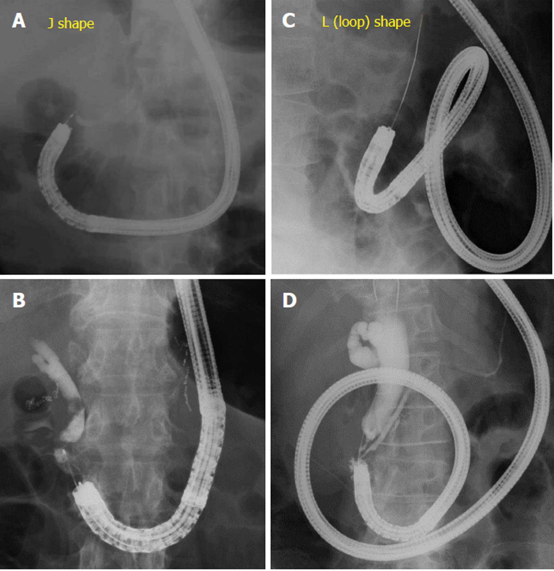
Figure 1 Shape of scope in Billroth-II reconstruction.
A, B: J-shaped scope inserted to the papilla; C, D: L-shaped, or looped, scope inserted to the papilla. The scope cannot be advanced to the papilla without looping the scope. Cases wherein looping is required during insertion, and releasing the loop during or after reaching the papilla, are included in this category.
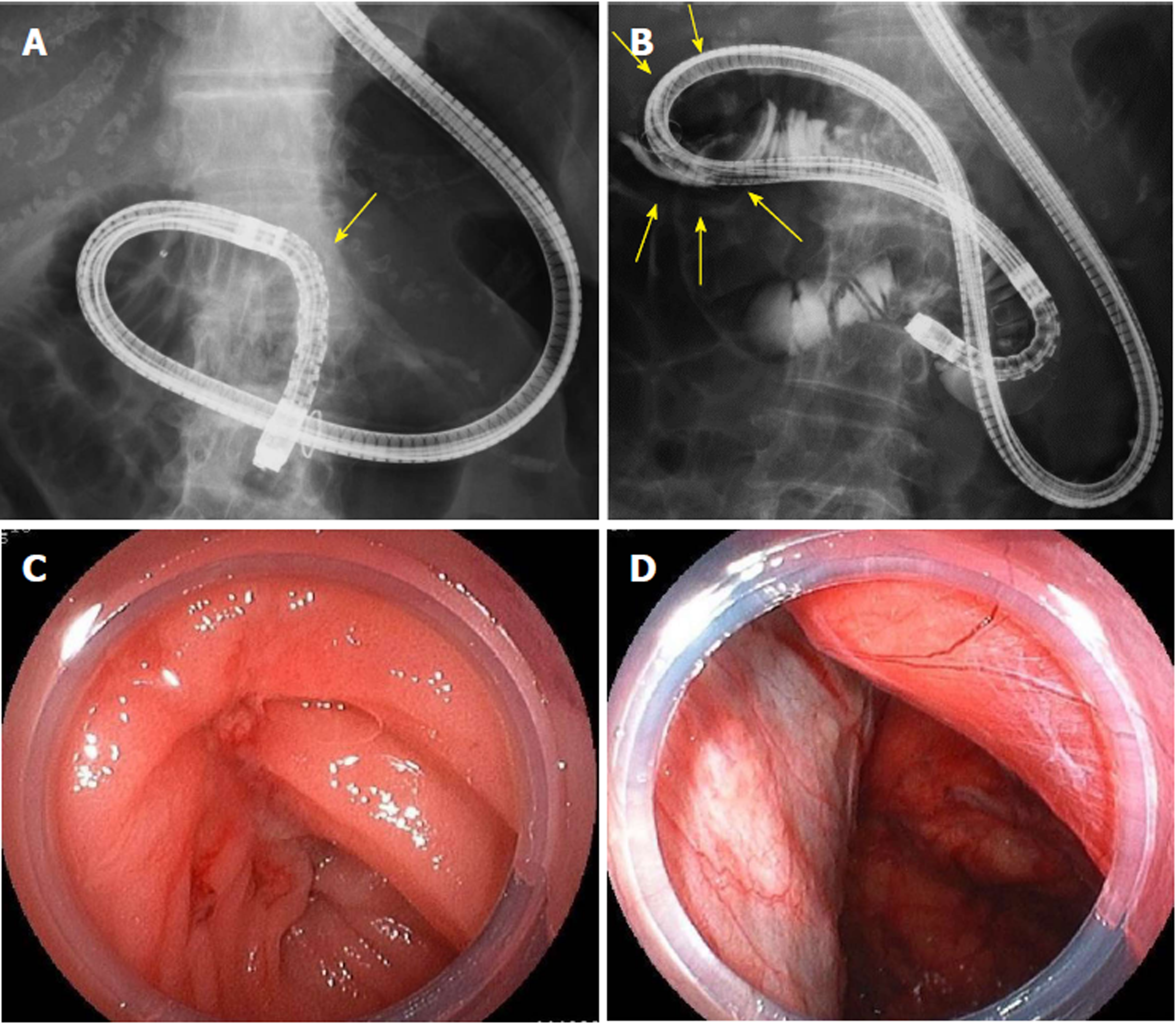
Figure 2 A case of gastrointestinal perforation during scope insertion after Billroth-II reconstruction.
A: Scope is being inserted retroflexed in the intestinal tract of Billroth-II anatomy. Arrow indicates the ligament of Treitz; B: Perforation of gastrointestinal tract occurred in an area showing air within retroperitoneal cavity (arrows); C: The area of perforation in gastrointestinal tract lumen; D: The retroperitoneal cavity seen from the area of perforation.
- Citation: Takano S, Fukasawa M, Shindo H, Takahashi E, Hirose S, Fukasawa Y, Kawakami S, Hayakawa H, Yokomichi H, Kadokura M, Sato T, Enomoto N. Risk factors for perforation during endoscopic retrograde cholangiopancreatography in post-reconstruction intestinal tract. World J Clin Cases 2019; 7(1): 10-18
- URL: https://www.wjgnet.com/2307-8960/full/v7/i1/10.htm
- DOI: https://dx.doi.org/10.12998/wjcc.v7.i1.10


