Copyright
©The Author(s) 2018.
World J Clin Cases. Dec 26, 2018; 6(16): 1182-1188
Published online Dec 26, 2018. doi: 10.12998/wjcc.v6.i16.1182
Published online Dec 26, 2018. doi: 10.12998/wjcc.v6.i16.1182
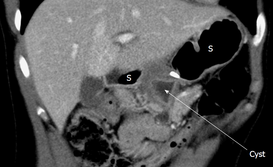
Figure 1 Computed tomography scan of the abdominal cavity performed because of persisting abdominal pain.
Peripancreatic thick-walled collection (cyst) located between the head of the pancreas and the posterior wall of the antrum of the stomach (S). In the context of the recent pancreatitis, the collection was initially interpreted as a pancreatic pseudocyst. Published with permission of Luzicka nemocnice a poliklinika, Rumburk.
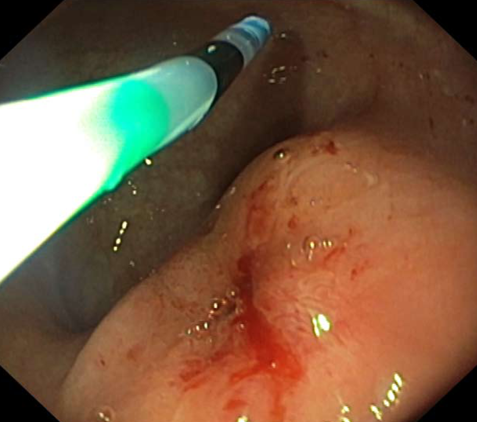
Figure 2 The gastroscopy shows a prominence of a posterior gastric wall.
It represents a gastric duplication cyst located at antrum of the stomach. Published with permission of Jablonec nad Nisou Hospital.
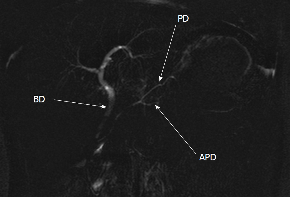
Figure 3 Magnetic resonance cholangiopancreatography shows the pancreatic duplication.
The accessory pancreatic duct of the accessory pancreatic lobe is located ventrally to the main pancreatic duct. The confluence is located in the body of the pancreas. The biliary duct is of common appearance. Published with permission of Jablonec nad Nisou Hospital. APD: Accessory pancreatic duct; PD: Pancreatic duct; BD: Biliary duct.
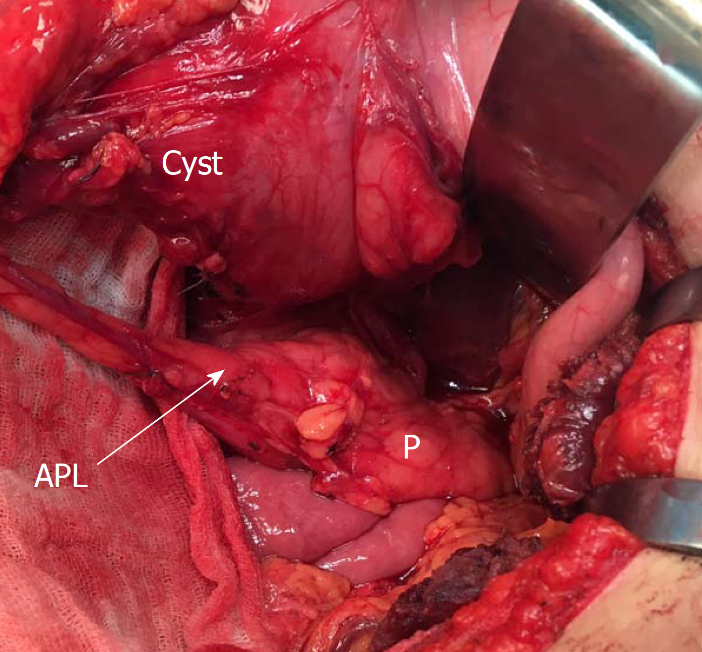
Figure 4 Perioperative finding of the gastric duplication cyst communicating to accessory pancreatic lobe.
The accessory pancreatic lobe arising from the body of the pancreas. It follows a ventral direction to the stomach and communicates with a gastric duplication cyst (cyst) located at the antrum of the stomach. APL: Accessory pancreatic lobe; P: Pancreas.
- Citation: Rousek M, Kachlik D, Nikov A, Pintova J, Ryska M. Gastric duplication cyst communicating to accessory pancreatic lobe: A case report and review of the literature. World J Clin Cases 2018; 6(16): 1182-1188
- URL: https://www.wjgnet.com/2307-8960/full/v6/i16/1182.htm
- DOI: https://dx.doi.org/10.12998/wjcc.v6.i16.1182


