Copyright
©The Author(s) 2018.
World J Clin Cases. Dec 26, 2018; 6(16): 1160-1163
Published online Dec 26, 2018. doi: 10.12998/wjcc.v6.i16.1160
Published online Dec 26, 2018. doi: 10.12998/wjcc.v6.i16.1160
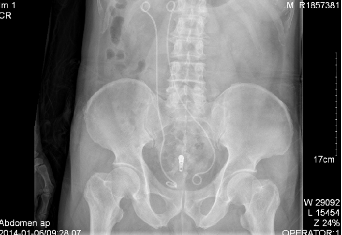
Figure 1 Plain kidney, ureter, and bladder X-ray showing the improperly positioned left ureteral stent.
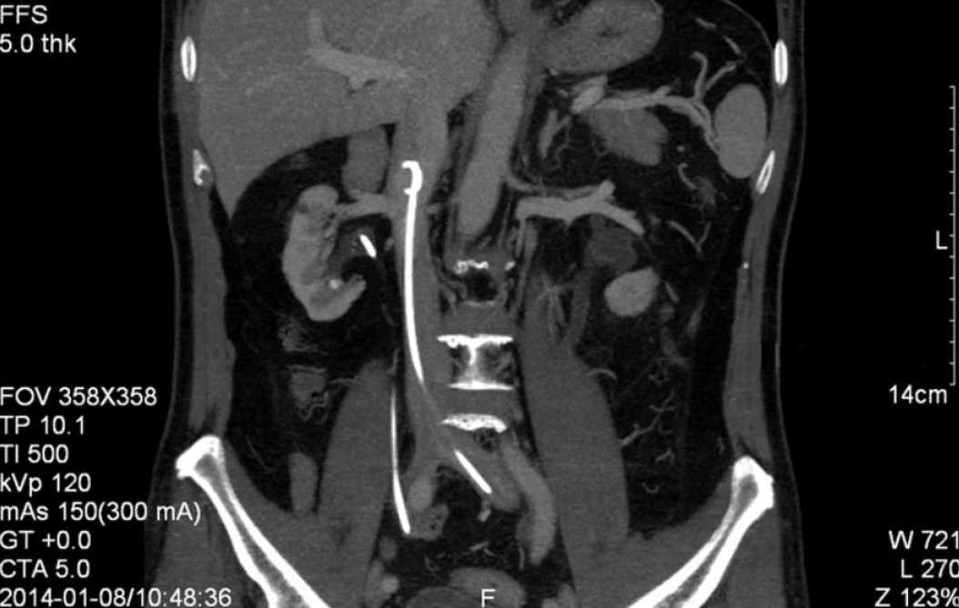
Figure 2 Computed tomography reconstruction of the abdomen confirmed the left ureteral stent in the inferior vena cava.
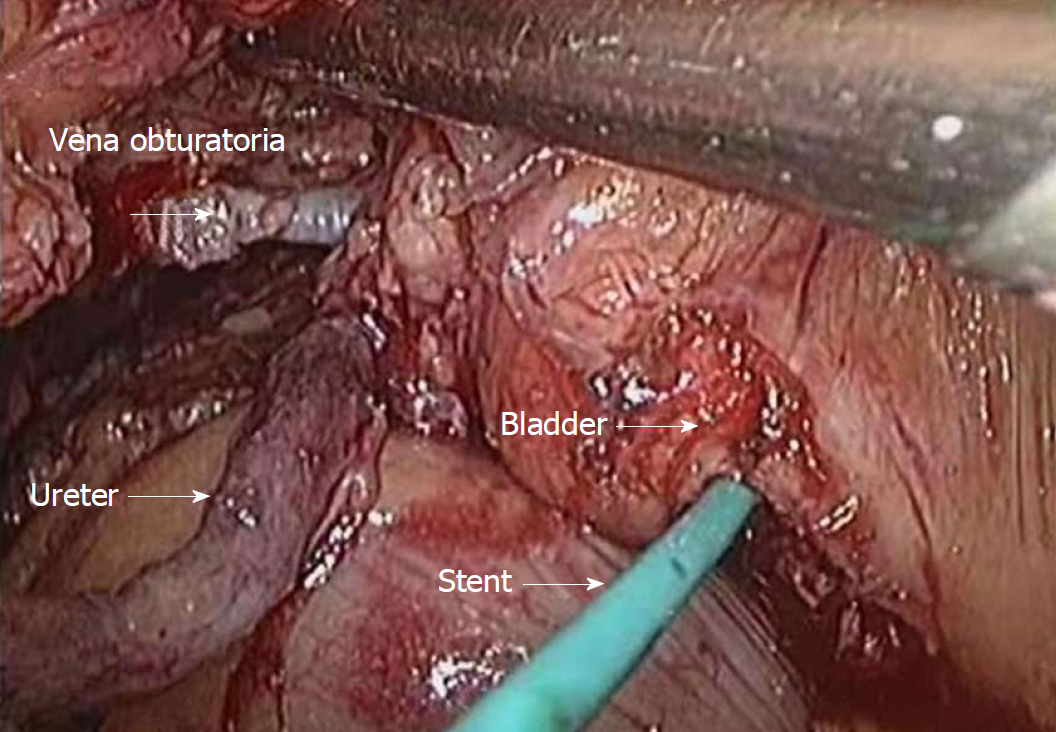
Figure 3 Surgical video screen capture demonstrating the corresponding anatomy.
- Citation: Mao XW, Xu G, Xiao JQ, Wu HF. Ureteral double J stent displaced into vena cava and management with laparoscopy: A case report and review of the literature. World J Clin Cases 2018; 6(16): 1160-1163
- URL: https://www.wjgnet.com/2307-8960/full/v6/i16/1160.htm
- DOI: https://dx.doi.org/10.12998/wjcc.v6.i16.1160


