Copyright
©The Author(s) 2018.
World J Clin Cases. Dec 6, 2018; 6(15): 1053-1058
Published online Dec 6, 2018. doi: 10.12998/wjcc.v6.i15.1053
Published online Dec 6, 2018. doi: 10.12998/wjcc.v6.i15.1053
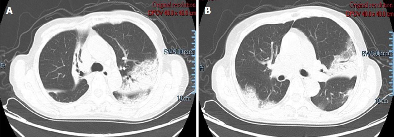
Figure 1 Chest computed tomography scan on admission showed bilateral lesions.
The main findings were patchy, consolidation and ground-glass opacities, associated with air bronchogram.
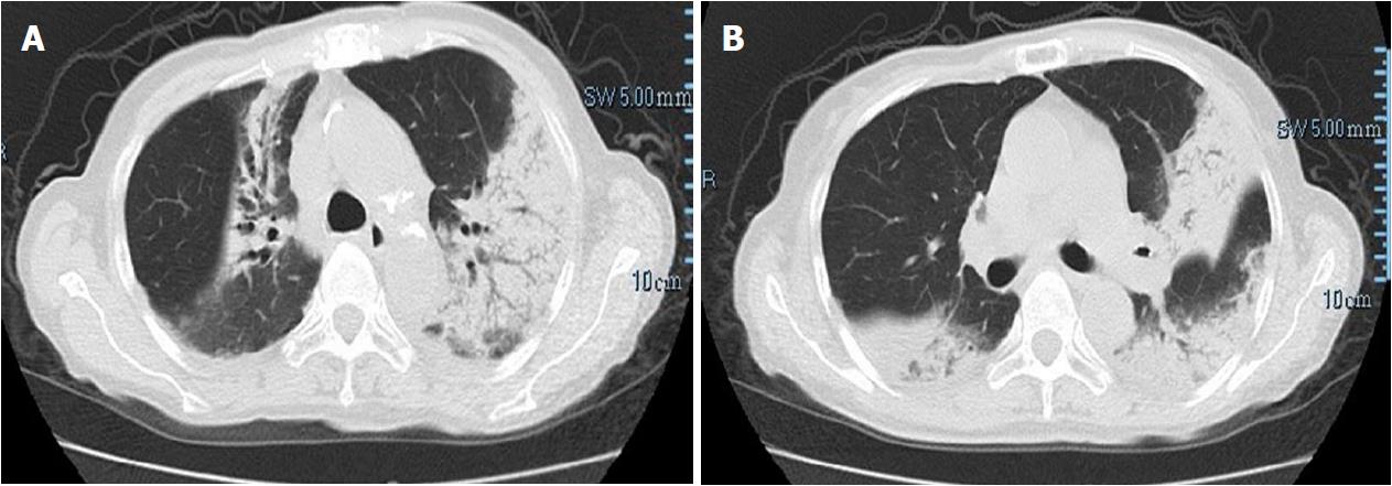
Figure 2 Repeat chest computed tomography scan showed an increase of the nodules and patchy infiltration after 2 wk antibacterial and antifungal treatment.
Dyspnea deteriorated and no improvement was observed.
Figure 3 Pathological examinations revealed numerous fibrin and organizing tissue in the alveoli without pulmonary hyaline membrane, the fibrous tissue hyperplasia in the alveolar septum, which were consistent with acute fibrinous and organizing pneumonia (original magnification × 400).
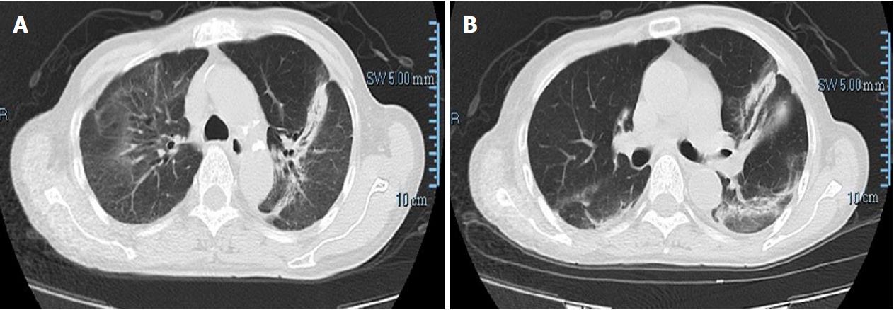
Figure 4 Repeat chest computed tomography scan after 1 wk steroid treatment showed significant improvement, and bilateral lesions resolved gradually.
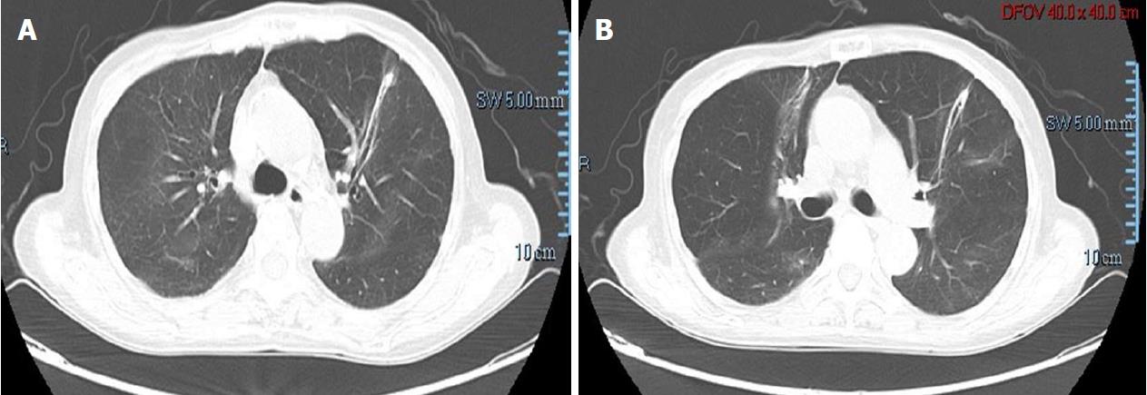
Figure 5 Repeat chest computed tomography scan after 3 wk steroid treatment showed significant improvement, and bilateral lesions almost disappeared.
- Citation: Ning YJ, Ding PS, Ke ZY, Zhang YB, Liu RY. Successful steroid treatment for acute fibrinous and organizing pneumonia: A case report. World J Clin Cases 2018; 6(15): 1053-1058
- URL: https://www.wjgnet.com/2307-8960/full/v6/i15/1053.htm
- DOI: https://dx.doi.org/10.12998/wjcc.v6.i15.1053


