Copyright
©The Author(s) 2018.
World J Clin Cases. Dec 6, 2018; 6(15): 1029-1035
Published online Dec 6, 2018. doi: 10.12998/wjcc.v6.i15.1029
Published online Dec 6, 2018. doi: 10.12998/wjcc.v6.i15.1029
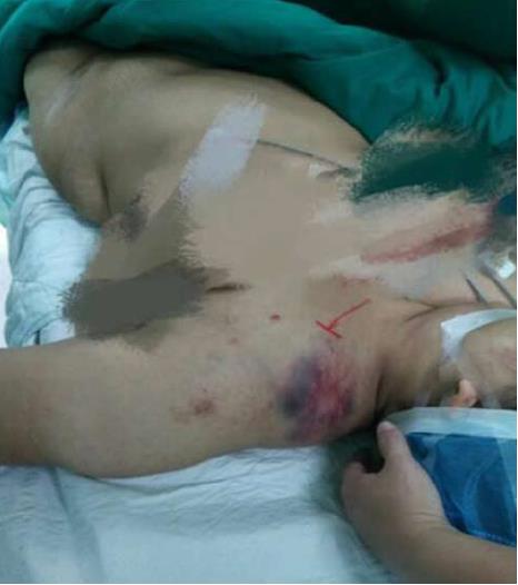
Figure 1 Closed left “floating shoulder”.
Marked soft tissue swelling, subcutaneous congestion, and deformity of the left shoulder were observed.
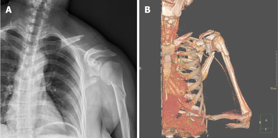
Figure 2 Plain anterior-posterior radiograph and computed tomography image of the left shoulder.
A: X-ray demonstrating fractures of the left scapula, left proximal humerus, left clavicle and left shoulder soft tissue swelling; B: 3D computed tomography angiography reconstruction of the left shoulder also demonstrating multiple fractures of the scapular region and interruption of blood flow through the axillary artery.
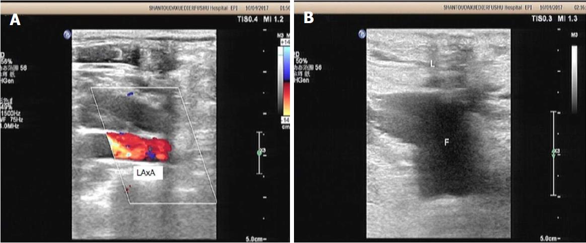
Figure 3 Doppler ultrasound revealed interruption of blood flow through the axillary artery.
A: The distal axillay artery was not unclear; B: The perivascular blood accumulation was obvious.
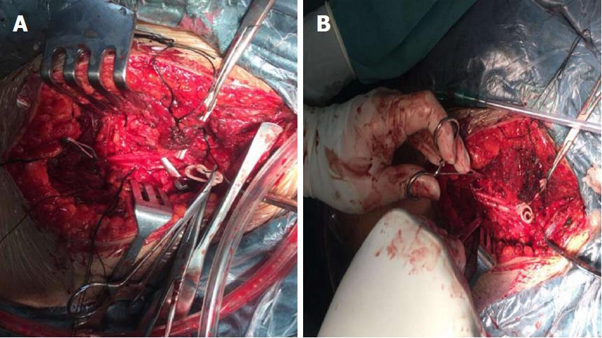
Figure 4 The axillary artery injury and repair.
A: Emergency surgery revealed that the axillary artery was completely amputated; B: End-to-end anastomoses were thus performed after excision of traumatized vessel segments.
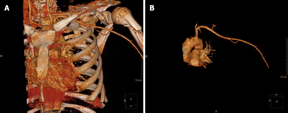
Figure 5 Evidence that axillary artery injury was successfully repaired.
A: Computed tomography (CT) angiography confirmed that blood flow of the left axillary artery was restored after vascular repair; B: CT angiography without bone confirmed that blood flow of the left axillary artery was restored after vascular repair.
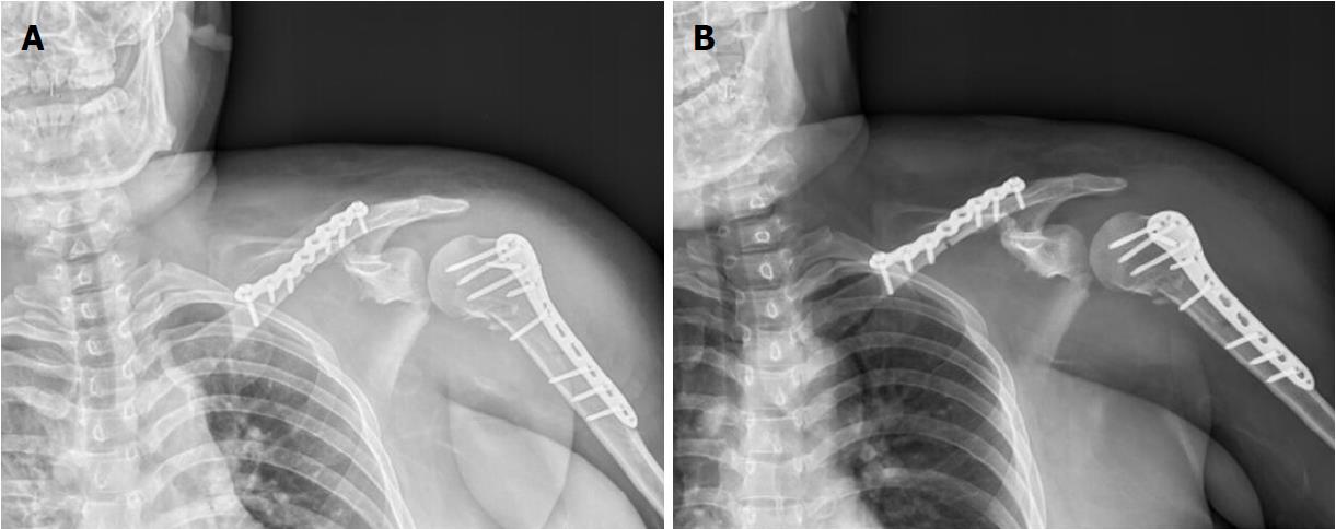
Figure 6 Postoperative radiographs showing reduction and locking plate.
A: Positive slice; B: Oblique slice.
- Citation: Chen YC, Lian Z, Lin YN, Wang XJ, Yao GF. Injury to the axillary artery and brachial plexus caused by a closed floating shoulder injury: A case report. World J Clin Cases 2018; 6(15): 1029-1035
- URL: https://www.wjgnet.com/2307-8960/full/v6/i15/1029.htm
- DOI: https://dx.doi.org/10.12998/wjcc.v6.i15.1029


