Copyright
©The Author(s) 2018.
World J Clin Cases. Dec 6, 2018; 6(15): 1007-1011
Published online Dec 6, 2018. doi: 10.12998/wjcc.v6.i15.1007
Published online Dec 6, 2018. doi: 10.12998/wjcc.v6.i15.1007
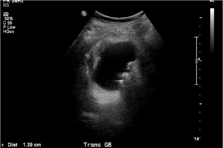
Figure 1 Ultrasound image demonstrating a pronounced gallbladder wall thickening and stones, consistent with acute colecystitis.
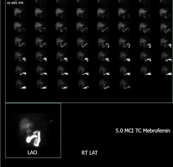
Figure 2 HIDA scan image demonstrates non-visualization of the gallbladder one hour after radiotracer injection, consistent with cystic duct obstruction.
- Citation: Mehrzad M, Jehle CC, Roussel LO, Mehrzad R. Gangrenous cholecystitis: A silent but potential fatal disease in patients with diabetic neuropathy. A case report. World J Clin Cases 2018; 6(15): 1007-1011
- URL: https://www.wjgnet.com/2307-8960/full/v6/i15/1007.htm
- DOI: https://dx.doi.org/10.12998/wjcc.v6.i15.1007


