Copyright
©The Author(s) 2016.
World J Clin Cases. Dec 16, 2016; 4(12): 390-400
Published online Dec 16, 2016. doi: 10.12998/wjcc.v4.i12.390
Published online Dec 16, 2016. doi: 10.12998/wjcc.v4.i12.390
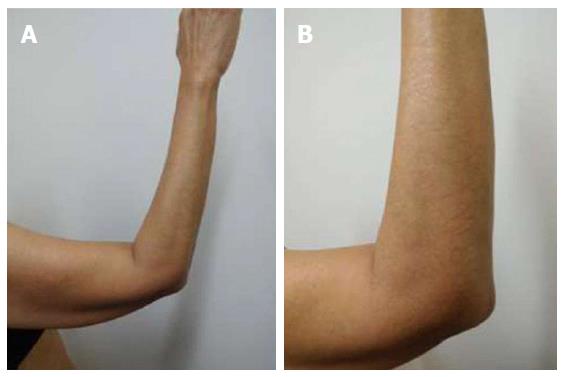
Figure 1 Distant (A) and closer (B) views of paclitaxel-induced reticulate hyperpigmentation on the elbow and extensor right arm.
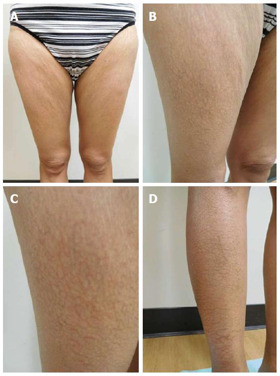
Figure 2 Paclitaxel-associated reticulate hyperpigmentation on the lower extremities.
Linear and net-like hyperpigmentation is noted on distant (A), intermediate (B and D) and closer (C) views of the right thigh (B and C) and right pretibial area (D).
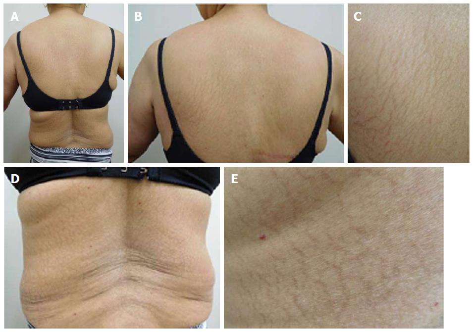
Figure 3 Paclitaxel-associated reticulate hyperpigmentation on the back.
Distant (A) view of the back shows reticulate hyperpigmentation following treatment with paclitaxel. Intermediate (B and D) and closer (C and E) views of the upper (B and C) and lower (D and E) back show the linear and net-like hyperpigmentation.
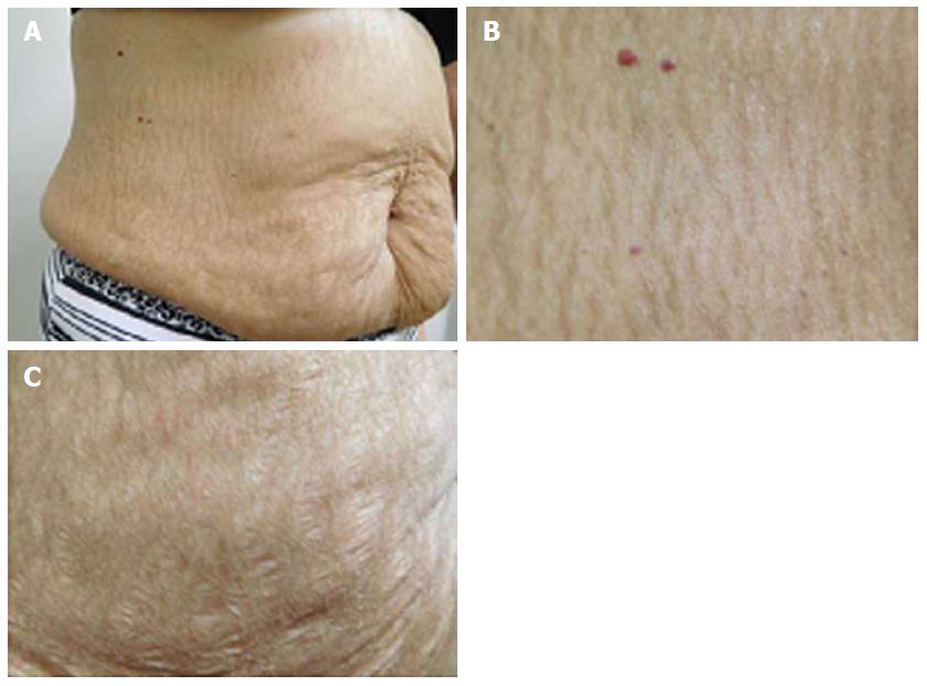
Figure 4 Paclitaxel-associated reticulate hyperpigmentation on the abdomen.
Distant (A) and closer (B) views of paclitaxel-induced reticulate hyperpigmentation on the abdomen. The hyperpigmentation spares the stretch marks on the abdomen (C).
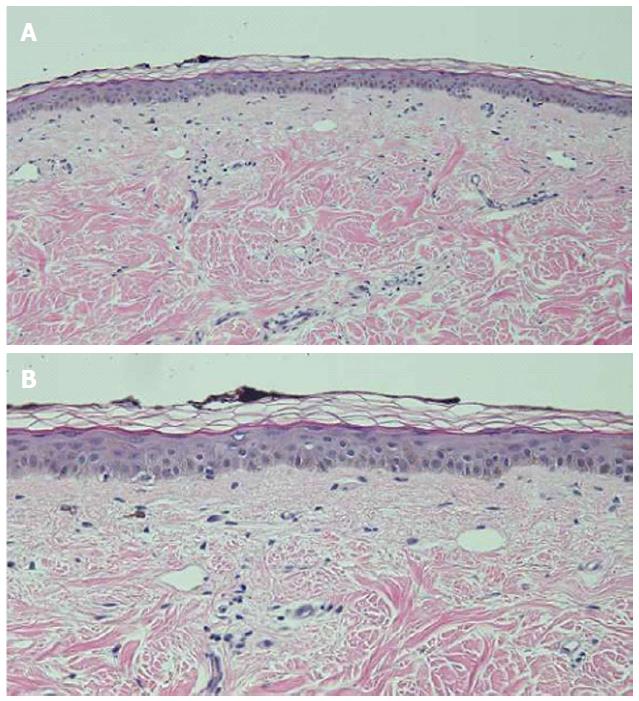
Figure 5 Distant (A) and closer (B) views of a hematoxylin and eosin stained biopsy specimen of the reticulate hyperpigmentation show confluent basilar hyperpigmentation with increased melanin in the basal layers of the epidermis.
Melanin is also present in melanophages in the papillary dermis (hematoxylin and eosin; A: × 10; B: × 20).
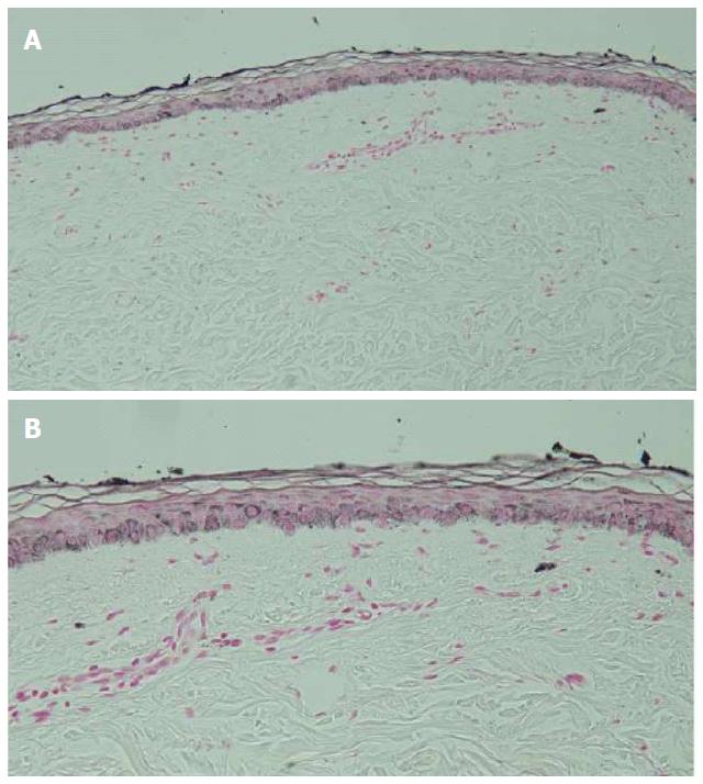
Figure 6 Distant (A) and closer (B) views of a Fontana-Masson stained biopsy specimen of the reticulate hyperpigmentation confirms the increased presence of melanin in the basal keratinocytes of the epidermis and within papillary dermis melanophages (Fontana-Masson; A: × 10; B: × 20).
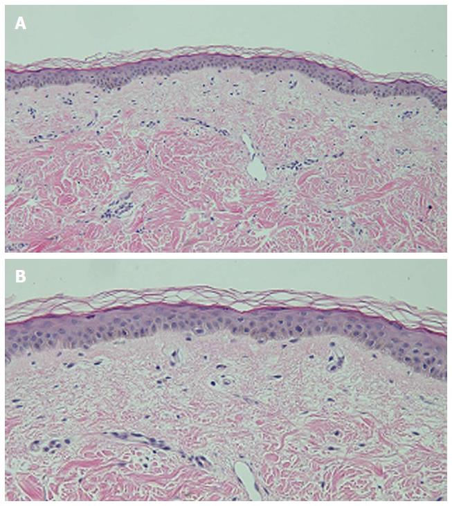
Figure 7 Distant (A) and closer (B) views of a hematoxylin and eosin stained biopsy specimen of normal appearing skin (adjacent to the hyperpigmentation) only shows minimal hyperpigmentation of the basal layer of the epidermis (hematoxylin and eosin; A: × 10; B: × 20).
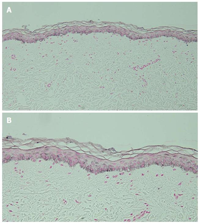
Figure 8 Distant (A) and closer (B) views of a Fontana-Masson stained biopsy specimen of normal appearing skin (adjacent to the hyperpigmentation) confirms the sparse presence of melanin in the epidermal basal layer and focally in the papillary dermis (Fontana-Masson; A: × 10; B: × 20).
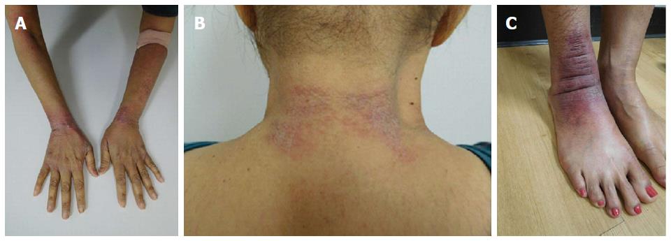
Figure 9 Paclitaxel-associated interface dermatitis presenting as tender and edematous red scaling plaques on the dorsal wrists (A), the posterior neck (B), and the right ankle (C).
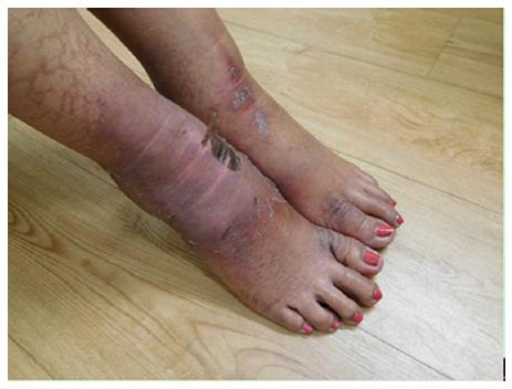
Figure 10 The paclitaxel-associated interface dermatitis lesion on the right ankle progressed in severity and distribution.
A similar lesion also developed on the left ankle.
- Citation: Cohen PR. Paclitaxel-associated reticulate hyperpigmentation: Report and review of chemotherapy-induced reticulate hyperpigmentation. World J Clin Cases 2016; 4(12): 390-400
- URL: https://www.wjgnet.com/2307-8960/full/v4/i12/390.htm
- DOI: https://dx.doi.org/10.12998/wjcc.v4.i12.390


