Copyright
©The Author(s) 2015.
World J Clin Cases. Aug 16, 2015; 3(8): 727-731
Published online Aug 16, 2015. doi: 10.12998/wjcc.v3.i8.727
Published online Aug 16, 2015. doi: 10.12998/wjcc.v3.i8.727
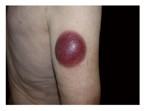
Figure 1 Right arm’s nodule, 6 cm × 6 cm in diameter.
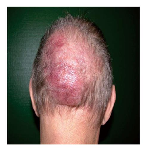
Figure 2 Vertex’s nodule, 16 cm × 13 cm in diameter.
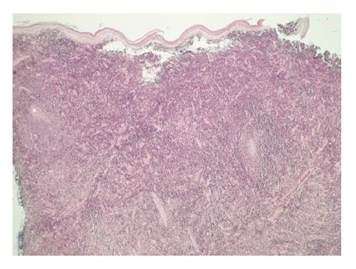
Figure 3 Histological aspect of a skin biopsy of the right arm’s nodule (HE).
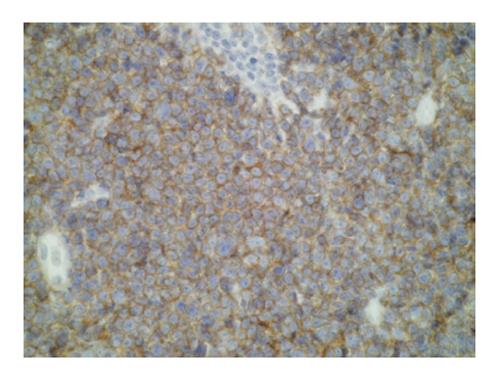
Figure 4 Tumour cells positive for CD4.
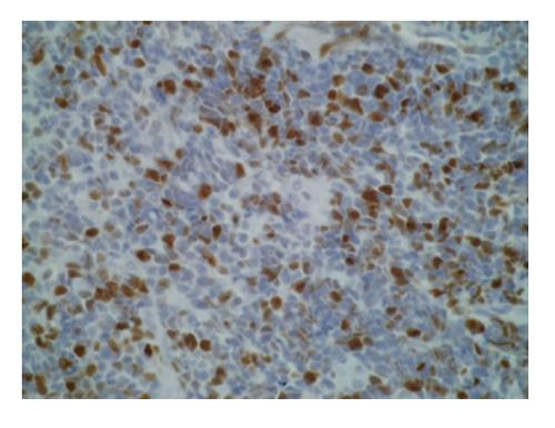
Figure 5 Tumour cells positive for ki67.
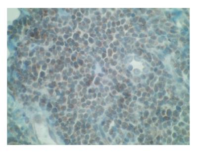
Figure 6 Positive terminal deoxynucleotidyl transferase marker.
- Citation: Ginoux E, Julia F, Balme B, Thomas L, Dalle S. T-lymphoblastic lymphoma with cutaneous involvement. World J Clin Cases 2015; 3(8): 727-731
- URL: https://www.wjgnet.com/2307-8960/full/v3/i8/727.htm
- DOI: https://dx.doi.org/10.12998/wjcc.v3.i8.727


