Copyright
©2014 Baishideng Publishing Group Inc.
World J Clin Cases. May 16, 2014; 2(5): 133-136
Published online May 16, 2014. doi: 10.12998/wjcc.v2.i5.133
Published online May 16, 2014. doi: 10.12998/wjcc.v2.i5.133
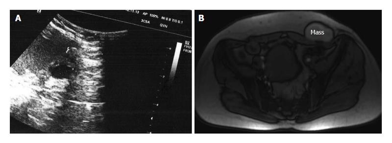
Figure 1 Ultrasonographic view (A) and magnetic resonance imaging (B) of scar endometrioma.
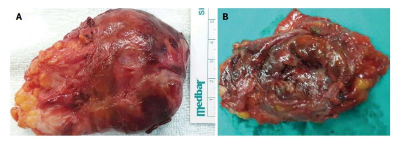
Figure 2 External view of resected endometrioma (A), cut surface of specimen showed semi-solid and soft tumor (B).
- Citation: Çöl C, Yilmaz EE. Cesarean scar endometrioma: Case series. World J Clin Cases 2014; 2(5): 133-136
- URL: https://www.wjgnet.com/2307-8960/full/v2/i5/133.htm
- DOI: https://dx.doi.org/10.12998/wjcc.v2.i5.133


