Copyright
©2014 Baishideng Publishing Group Co.
World J Clin Cases. Apr 16, 2014; 2(4): 100-103
Published online Apr 16, 2014. doi: 10.12998/wjcc.v2.i4.100
Published online Apr 16, 2014. doi: 10.12998/wjcc.v2.i4.100
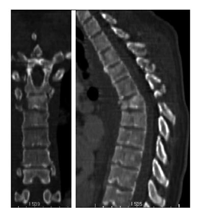
Figure 1 Severe rotation injury of T6 in the coronal and sagittal plane.
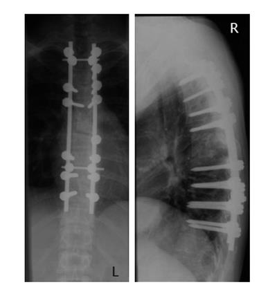
Figure 2 Postoperative X-ray after posterior fusion.
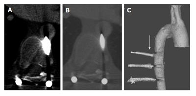
Figure 3 The left pedicle screw of T7 seems to perforate the aorta.
A and B: Transversal view; C: 3D image, the left T7 pedicle screw is marked by the arrow.
- Citation: Decker S, Omar M, Krettek C, Müller CW. Elective thoracotomy for pedicle screw removal to prevent severe aortic bleeding. World J Clin Cases 2014; 2(4): 100-103
- URL: https://www.wjgnet.com/2307-8960/full/v2/i4/100.htm
- DOI: https://dx.doi.org/10.12998/wjcc.v2.i4.100


