Copyright
©2014 Baishideng Publishing Group Inc.
World J Clin Cases. Oct 16, 2014; 2(10): 522-527
Published online Oct 16, 2014. doi: 10.12998/wjcc.v2.i10.522
Published online Oct 16, 2014. doi: 10.12998/wjcc.v2.i10.522
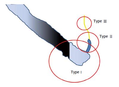
Figure 1 Classification of endoscopic retrograde cholangiopancreatography-related perforation (based on Kim et al[14]).
Figure 2 Endoscopic visualization of perforations.
A: Image of yellowish tissue in the retroperitoneum; B and C: Bleeding from a lateral wall of the duodenum.
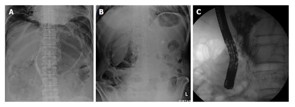
Figure 3 Fluoroscopy showing pneumoperitoneum (A), retroperitoneal air (B) and leakage of contrast media into the retroperitoneal cavity (C).
The kidney outline (clear region on the right side of the image) showing retroperitoneal air.
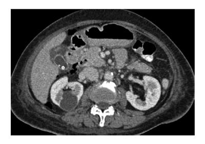
Figure 4 Computed tomography showing retroperitoneal air.
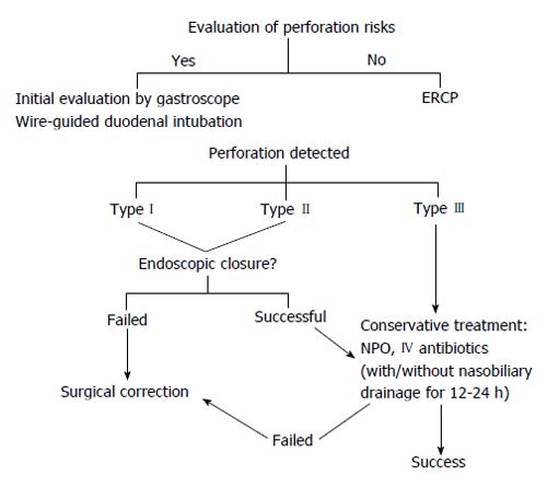
Figure 5 Algorithm for prevention and management of endoscopic retrograde cholangiopancreatography-related perforations.
- Citation: Prachayakul V, Aswakul P. Endoscopic retrograde cholangiopancreatography-related perforation: Management and prevention. World J Clin Cases 2014; 2(10): 522-527
- URL: https://www.wjgnet.com/2307-8960/full/v2/i10/522.htm
- DOI: https://dx.doi.org/10.12998/wjcc.v2.i10.522


