
















Copyright: ©Author(s) 2026.
World J Clin Cases. Sep 6, 2026; 14(25): 123670
Published online Sep 6, 2026. doi: 10.12998/wjcc.123670
Published online Sep 6, 2026. doi: 10.12998/wjcc.123670
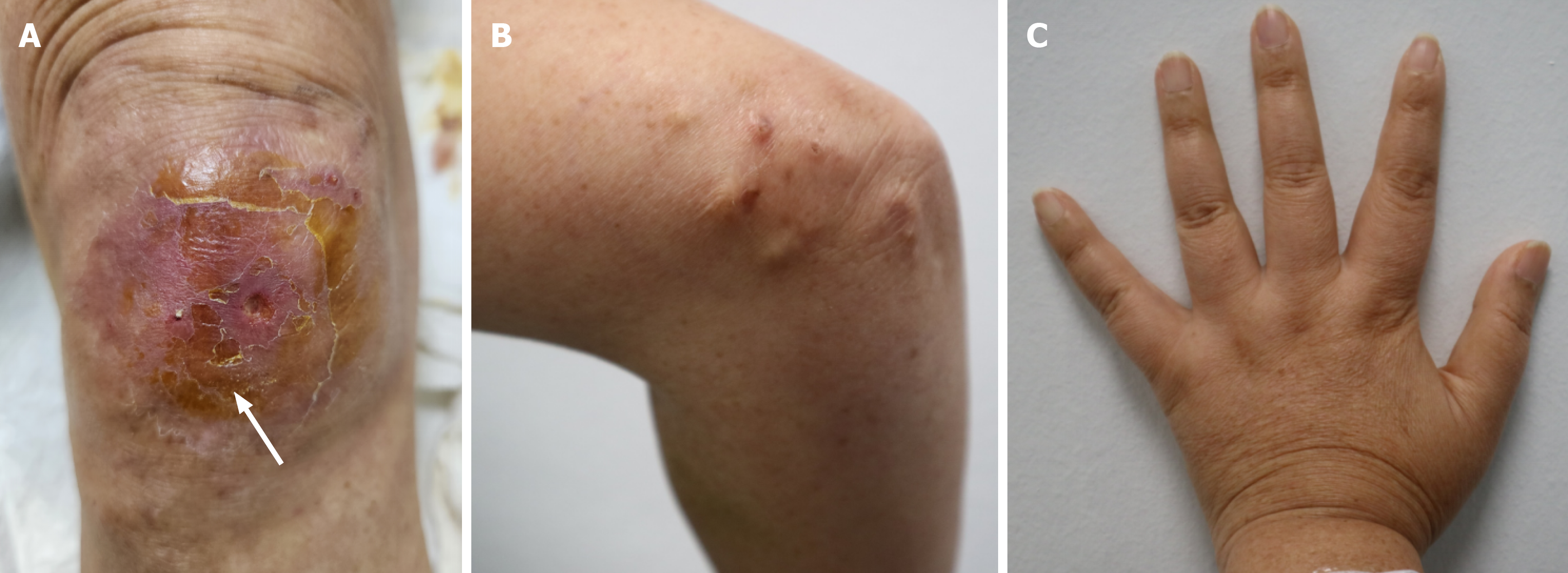
Figure 1 Gross photographs of skin lesions in a 59-year-old woman.
Multiple calcinosis cutis observed. A: Right knee; white arrow indicates skin ulceration with draining serous discharge; B: Right elbow; C: Left hand showing clinically diagnosed sclerodactyly, with thickened, waxy skin.
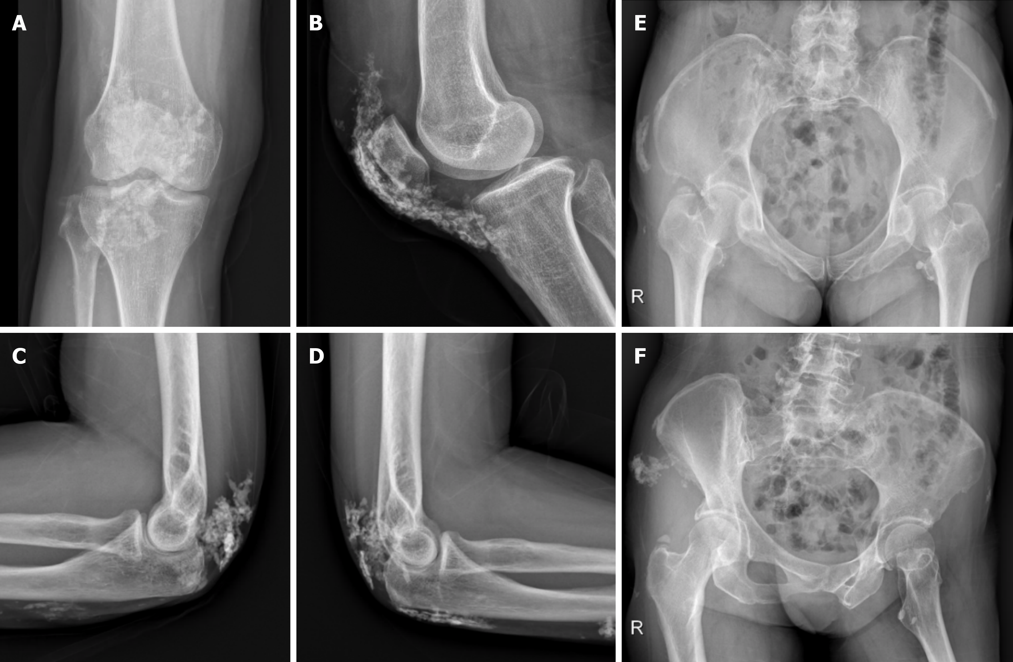
Figure 2 Preoperative simple radiographs displaying multiple, large-sized calcinosis cutis at various unusual locations, with no evidence of malignancy or infectious conditions detected.
A and B: Anteroposterior and lateral views of the right knee, respectively; C and D: Lateral views of the right and left elbows, respectively; E and F: Pelvic inlet view and right oblique view, respectively.
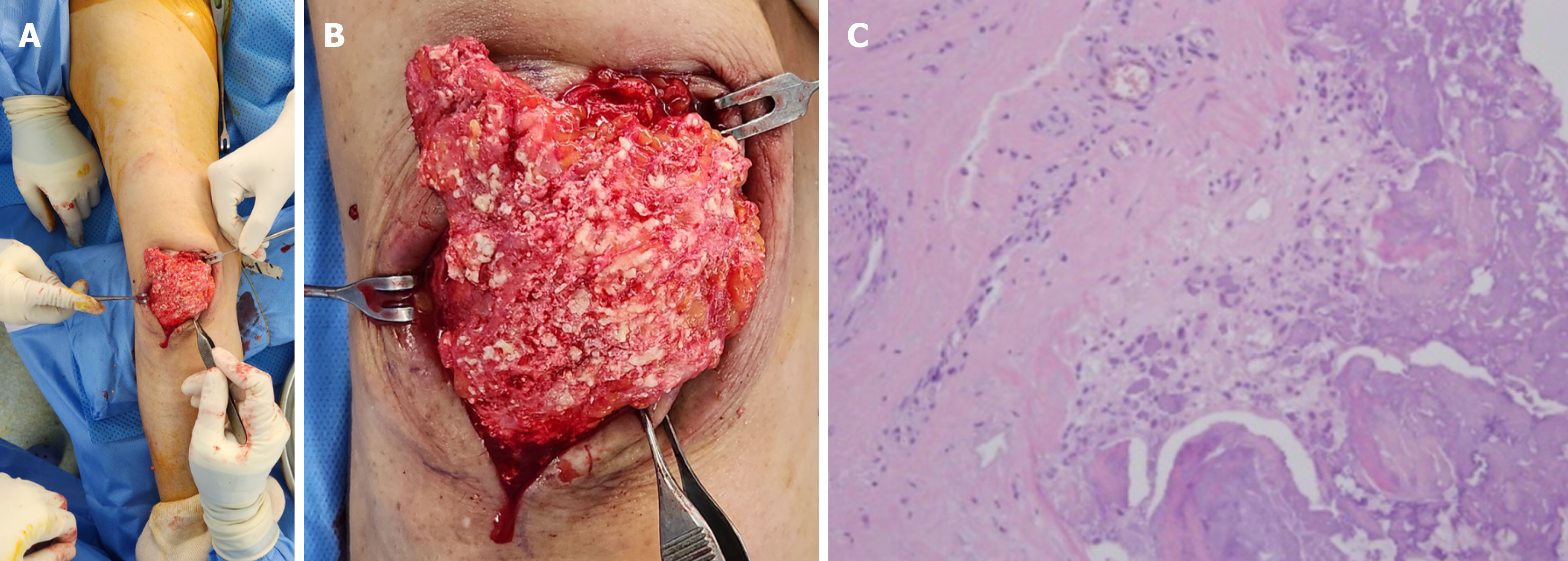
Figure 3 Excisional biopsy procedure.
A: Careful dissection was performed between the thinned skin tissue and subcutaneous calcified tissue. B: The extracted calcified tissue. C: Permanent biopsy analysis under an optical microscope (200× magnification, hematoxylin and eosin stain) revealed aggregated lymphocytes and fibrosis around the massive calcification, suggestive of an autoimmune response.
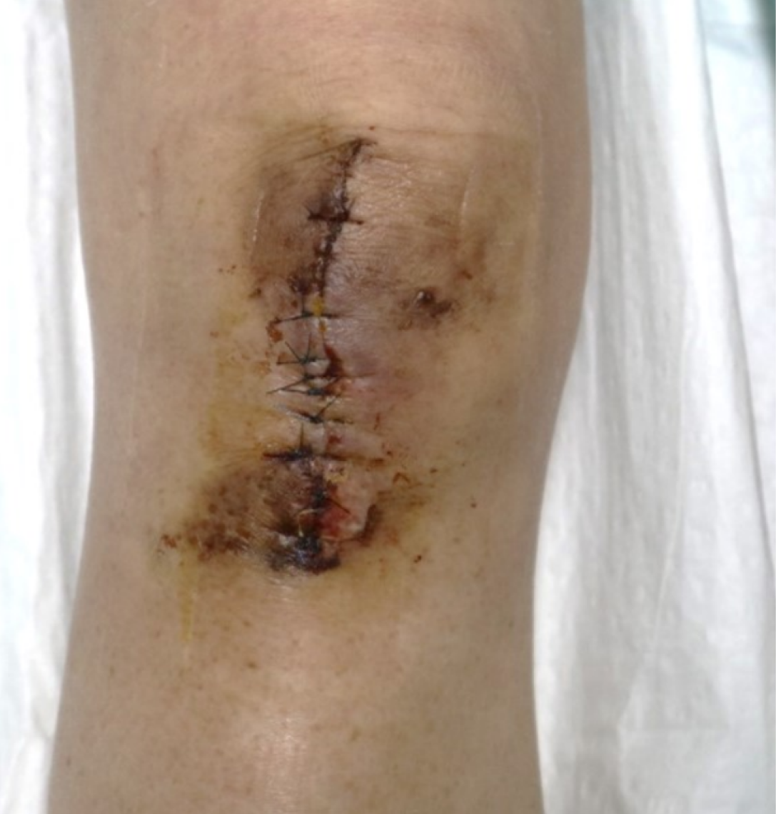
Figure 4 Surgical wound at two weeks postoperative, showing total stitch removal despite focal skin necrosis at the wound margin.
The wound spontaneously healed by the four-week postoperative follow-up visit.
- Citation: Lee HY, Lee SK, Kim H, Kang SW. Extensive multiple calcinosis cutis in seronegative limited cutaneous scleroderma (CREST variant): A case report. World J Clin Cases 2026; 14(25): 123670
- URL: https://www.wjgnet.com/2307-8960/full/v14/i25/123670.htm
- DOI: https://dx.doi.org/10.12998/wjcc.123670