Copyright
©The Author(s) 2025.
World J Clin Cases. Oct 16, 2025; 13(29): 108582
Published online Oct 16, 2025. doi: 10.12998/wjcc.v13.i29.108582
Published online Oct 16, 2025. doi: 10.12998/wjcc.v13.i29.108582
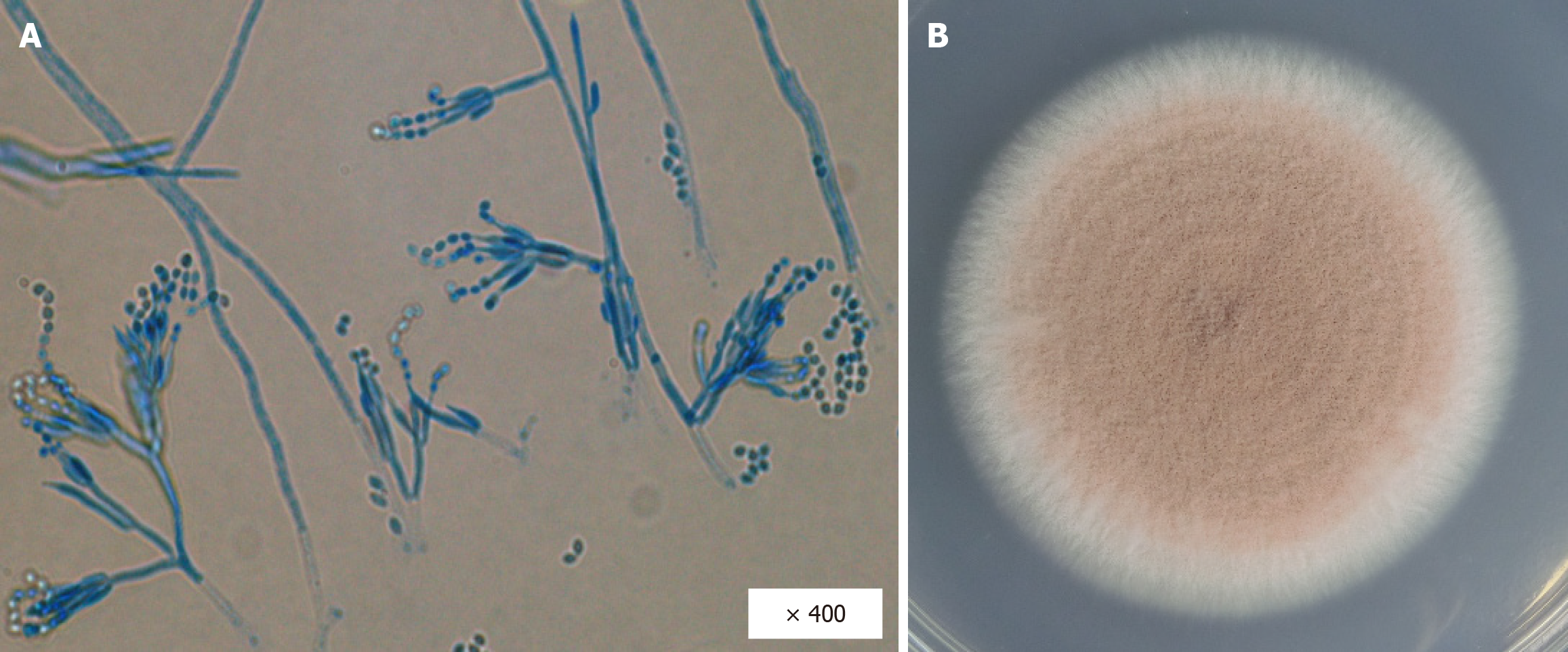
Figure 1 Morphological findings of the direct sputum smear.
A: Microscopic findings with lactophenol cotton blue staining (magnification: 400 ×), and numerous branching mycelia with septa, together with broom-like conidiogenous cells associated with oval condia, are confirmed; B: Light purple colonies with velvety surface are grown on potato dextrose agar.
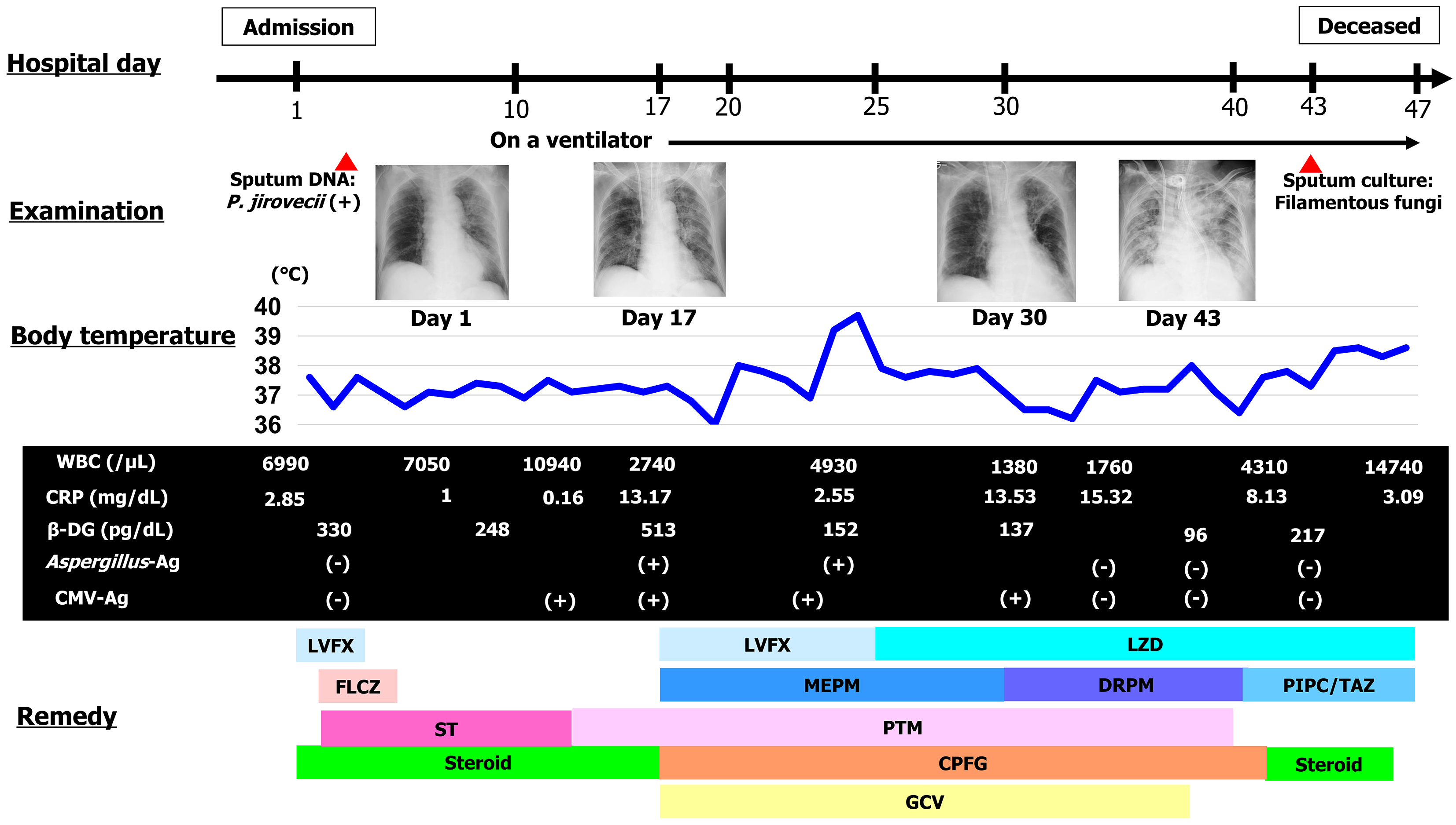
Figure 2 The clinical course of our case.
Ag: Antigen; β-DG: Beta-D-glucan; CMV: Cytomegalovirus; CPFG: Caspofungin; CRP: C-reactive protein; DNA: Deoxyribonucleic acid; DRPM: Doripenem; FLCZ: Fluconazole; GCV: Ganciclovir; LVFX: Levofloxacin; LZD: Linezolid; MEPM: Meropenem; PIPC/TAZ: Piperacillin/tazobactam; P. jirovecii: Pneumocystis jirovecii; PTM: Pentamidine; ST: Sulfamethoxazole and trimethoprim; WBC: White blood cell.
- Citation: Usuda D, Furukawa D, Imaizumi R, Ono R, Kaneoka Y, Nakajima E, Sugawara Y, Shimizu R, Sakurai R, Matsubara S, Tanaka R, Suzuki M, Shimozawa S, Hotchi Y, Osugi I, Katou R, Ito S, Mishima K, Kondo A, Mizuno K, Takami H, Komatsu T, Nomura T, Sugita M. Purpureocillium lilacinum: A minireview. World J Clin Cases 2025; 13(29): 108582
- URL: https://www.wjgnet.com/2307-8960/full/v13/i29/108582.htm
- DOI: https://dx.doi.org/10.12998/wjcc.v13.i29.108582


