Copyright
©The Author(s) 2025.
World J Clin Cases. Sep 16, 2025; 13(26): 108409
Published online Sep 16, 2025. doi: 10.12998/wjcc.v13.i26.108409
Published online Sep 16, 2025. doi: 10.12998/wjcc.v13.i26.108409
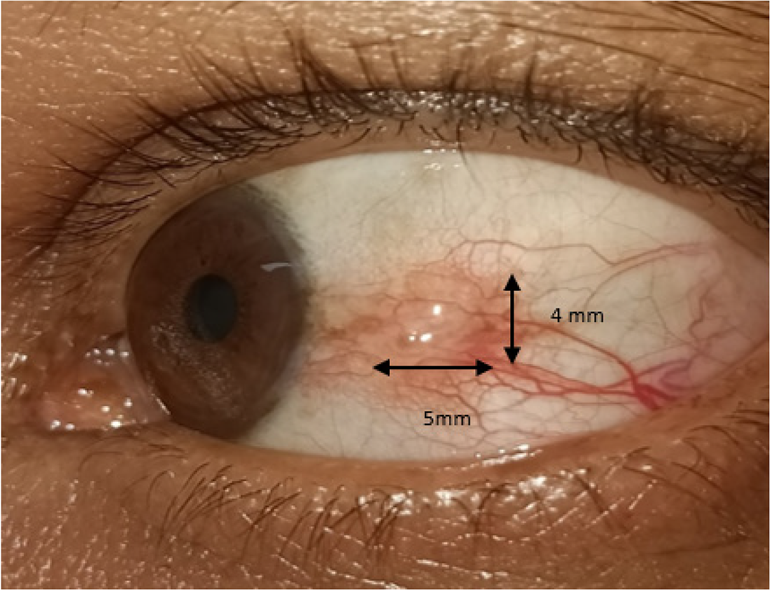
Figure 1
Clinical photo of left eye conjunctival mass size 5 mm × 4 mm extending from limbus with prominent intrinsic vascularity suggestive of ocular surface squamous neoplasia.
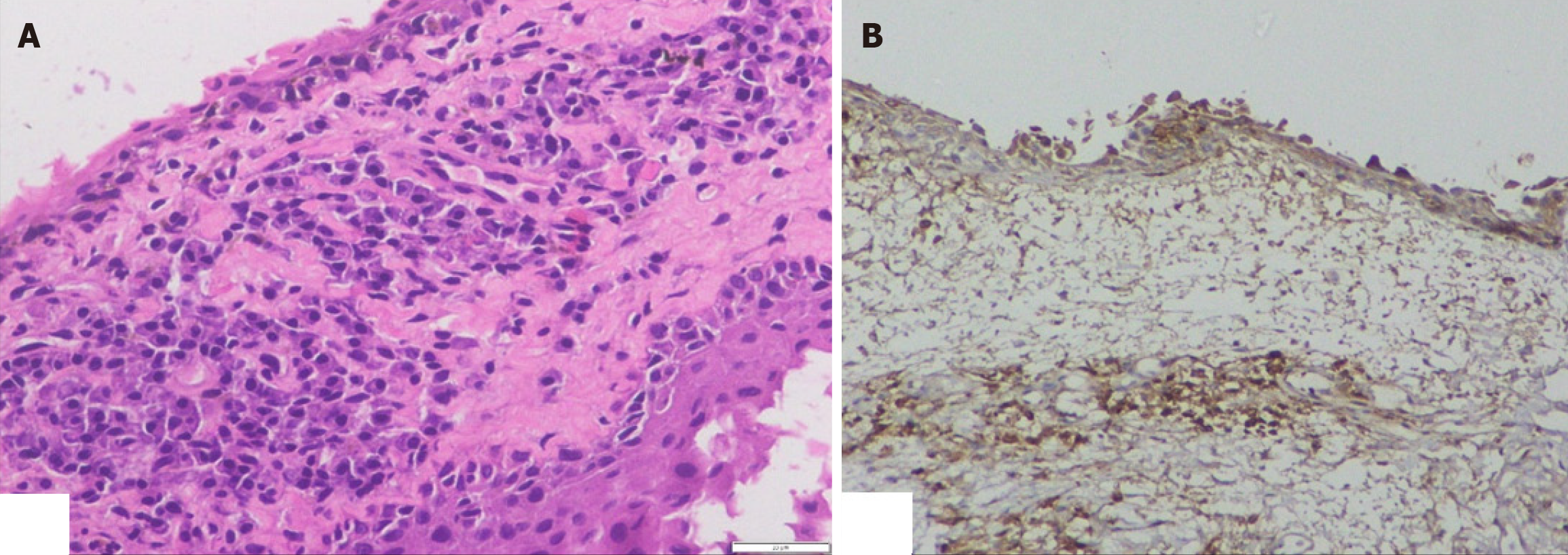
Figure 2 Histopathological changes of conjunctiva showing.
A: Sub conjunctival perivascular aggregates of reactive plasma cells (H&E, 40×); B: Immunohistochemistry (20×) of conjunctival mass showing plasma cells immune-positive for CD138, kappa, and lambda.
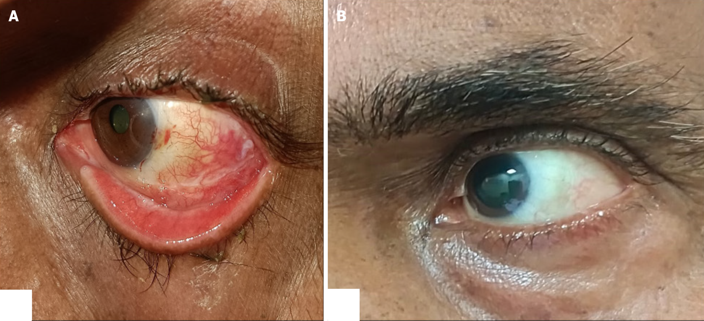
Figure 3 Clinical photo at follow up of two weeks and six months respectively showing.
A: Amniotic membrane in place with minimal congestion and sub conjunctival hemorrhage; B: Healed conjunctiva without any recurrence.
- Citation: Koppalu Lingaraju T, Panda BB, Sethy M, Achanta SL. Solitary extramedullary plasmacytoma mimicking ocular surface squamous neoplasia in an elderly male: A case report. World J Clin Cases 2025; 13(26): 108409
- URL: https://www.wjgnet.com/2307-8960/full/v13/i26/108409.htm
- DOI: https://dx.doi.org/10.12998/wjcc.v13.i26.108409


