Copyright
©The Author(s) 2025.
World J Clin Cases. Sep 16, 2025; 13(26): 106999
Published online Sep 16, 2025. doi: 10.12998/wjcc.v13.i26.106999
Published online Sep 16, 2025. doi: 10.12998/wjcc.v13.i26.106999
Figure 1 Preoperative magnetic resonance imaging showing a very large, rounded mass was observed in the pelvis, with clear borders and a visible periphery.
A and B: Representative longitudinal sections; C: Transverse section.
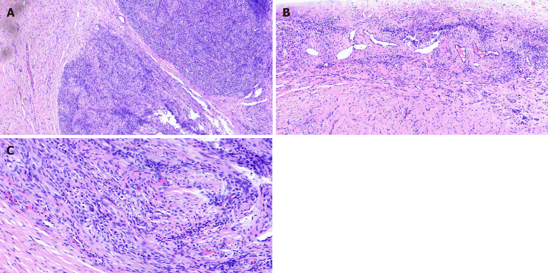
Figure 2 Postoperative hematoxylin and eosin staining pathology findings consistent with ovarian sclerosing mesenchymal tumors.
A: Magnification (100 ×); B: Magnification (200 ×); C: Magnification (400 ×).
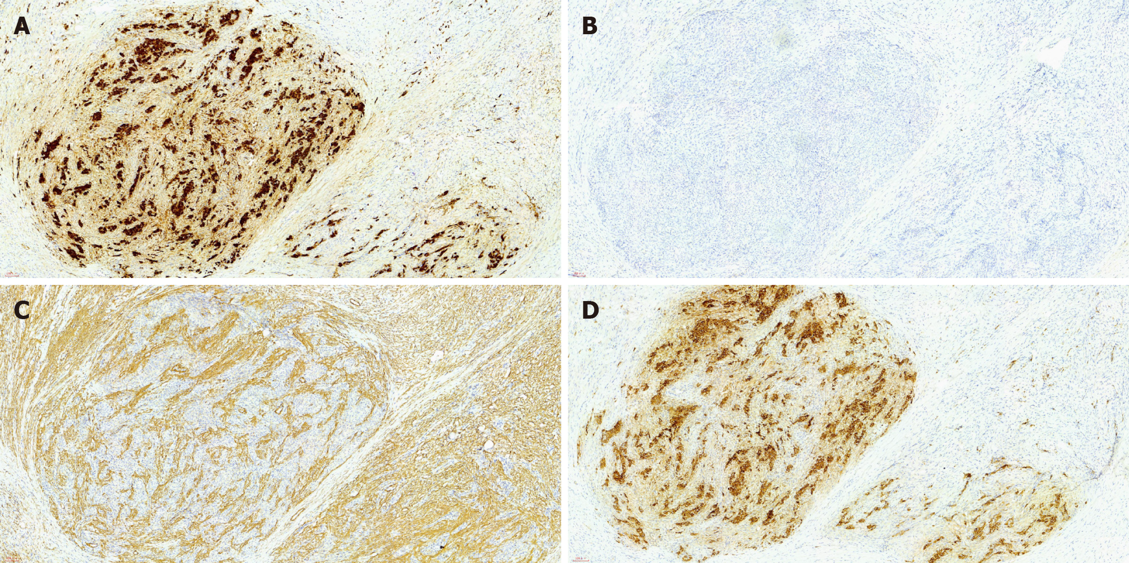
Figure 3 Immunohistochemical staining of the pathological sections.
A: Calretinin (+); B: Epithelial membrane antigen (-); C: Smooth muscle actin (partial +); D: Α-inhibin (+). Images were taken at magnification (100 ×).
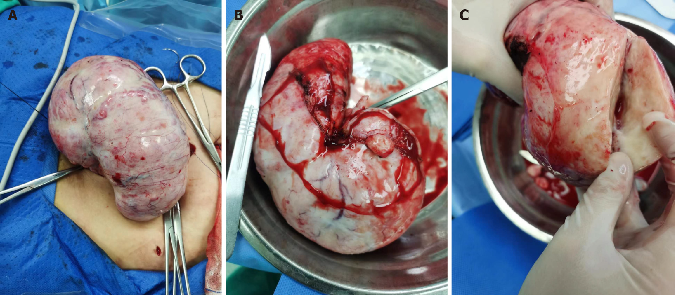
Figure 4 Intraoperative section of the ovarian.
A and B: Gross view of the ovarian mass, showing a huge solid mass, grayish white in color, textured, and hard and very rich in surface blood vessels; C: Section of the left ovarian mass.
- Citation: Han X, Zhang B, Gao JC. Late menstruation with an ovarian sclerosing stromal tumor: A case report. World J Clin Cases 2025; 13(26): 106999
- URL: https://www.wjgnet.com/2307-8960/full/v13/i26/106999.htm
- DOI: https://dx.doi.org/10.12998/wjcc.v13.i26.106999


