Copyright
©The Author(s) 2024.
World J Clin Cases. Feb 26, 2024; 12(6): 1111-1119
Published online Feb 26, 2024. doi: 10.12998/wjcc.v12.i6.1111
Published online Feb 26, 2024. doi: 10.12998/wjcc.v12.i6.1111
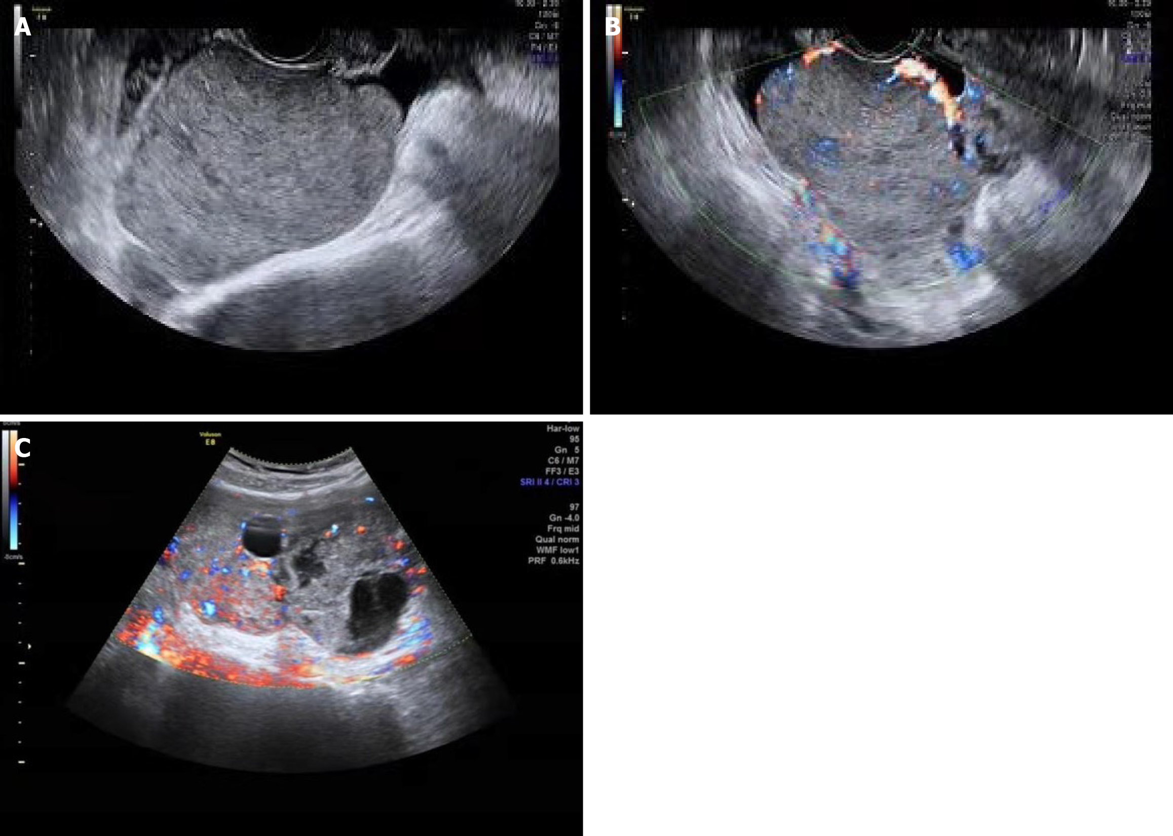
Figure 1 Ultrasound features.
A: The tumor was solid with heterogeneous echo in case 4; B and C: Color Doppler flow imaging displayed rich intralesional vascularization in the solid part of the lesion.
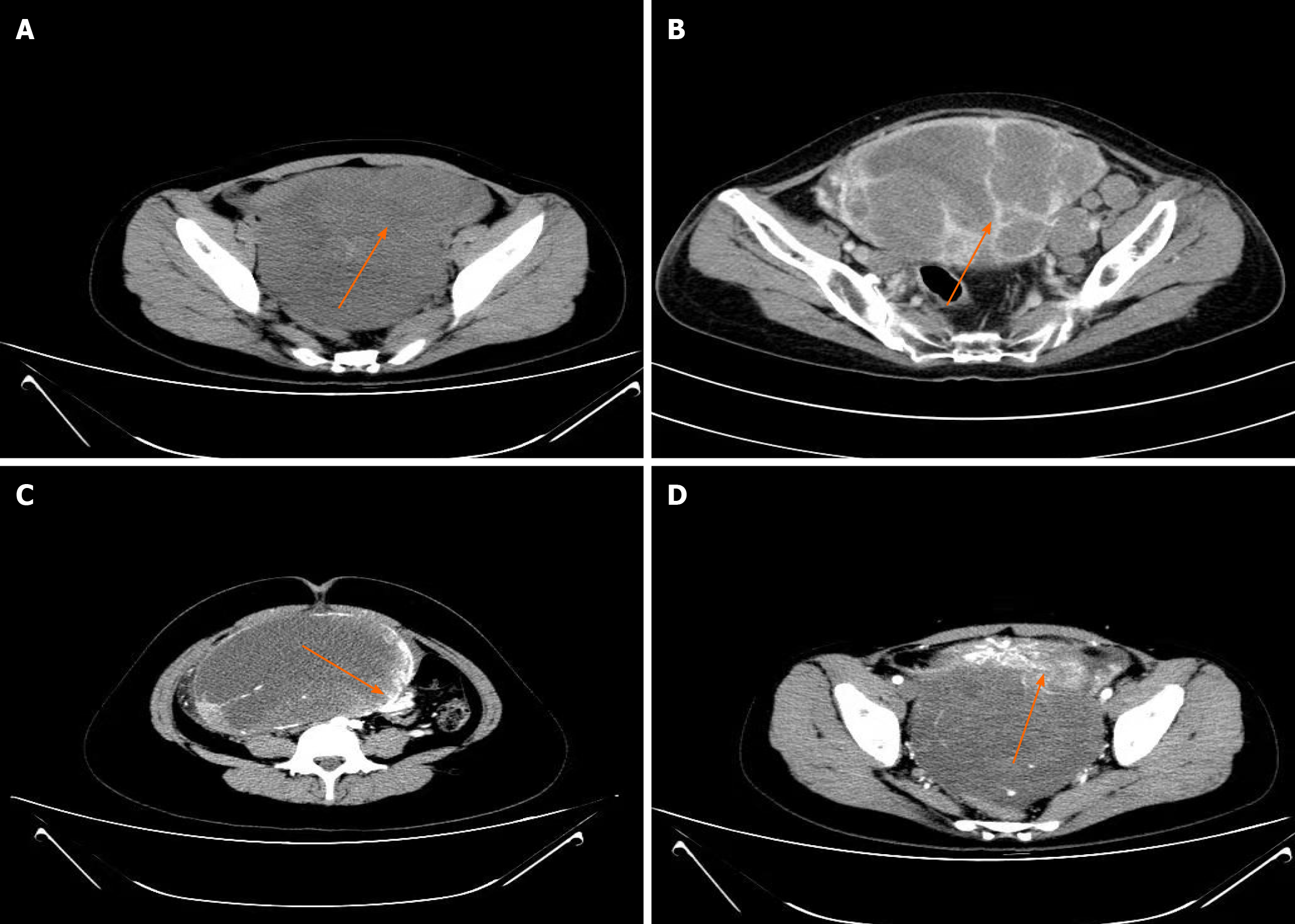
Figure 2 Computed tomography features.
A: A soft tissue mass with heterogeneous density occupied the pelvic cavity in case 1; B: Computed tomography (CT) scan revealed a multiseptated mixed solid and cystic mass in case 12; C: Enhanced CT showed marginal enhancement of the tumor in case 5; D: Heterogenous enhancement of the solid part.
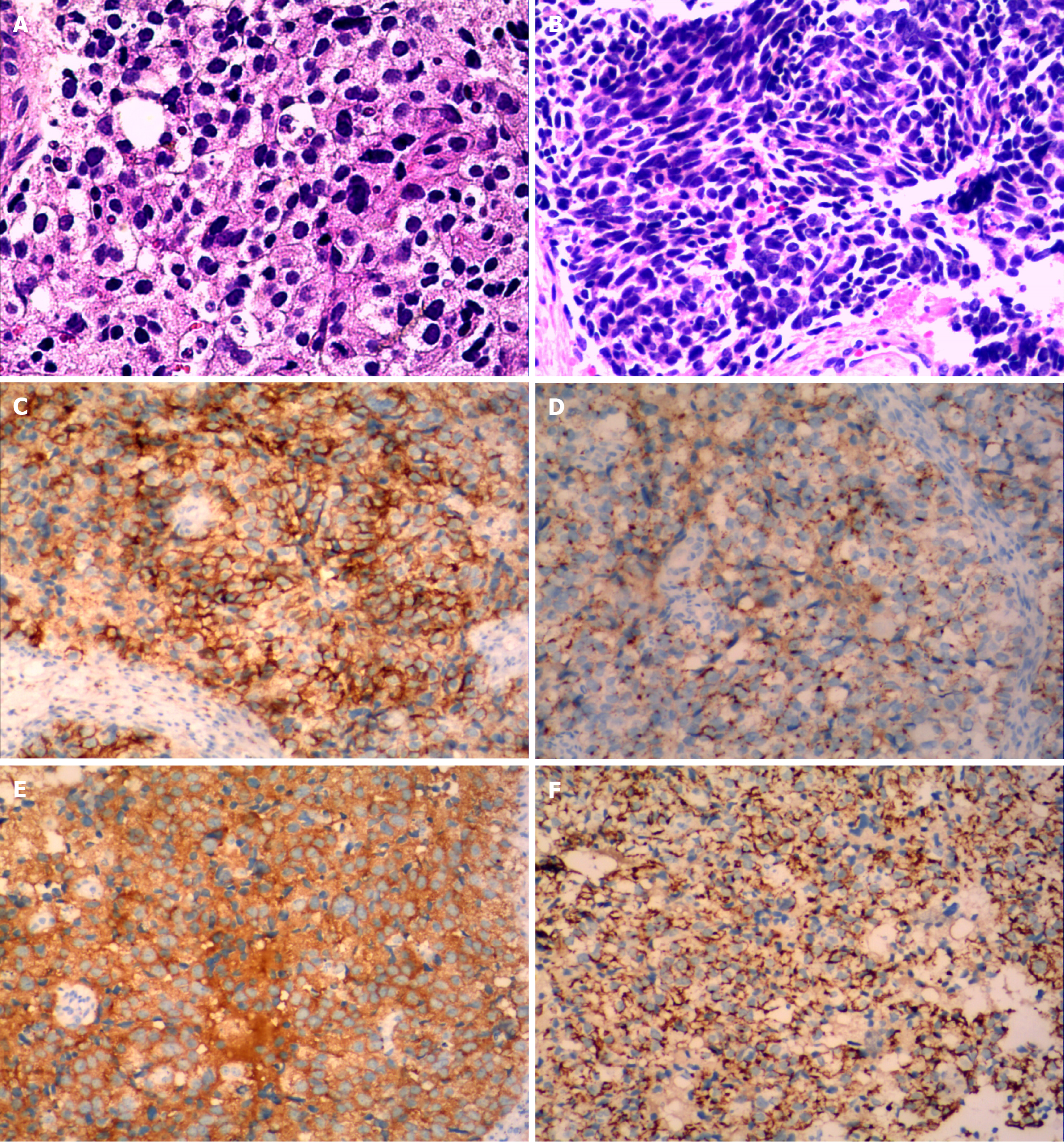
Figure 3 Histological and immunohistochemical large cell neuroendocrine carcinoma.
A: Tumor cells are arranged in nests, with relatively abundant basophilic cytoplasm, large round-to-oval nuclei and frequent mitosis in case 10; B: Small cell carcinoma of the ovary: Tumor cells are arranged in an organoid pattern, and some of the cells are spindle-shaped. The nucleus is oat-like, with abundant mitotic and apoptotic activity in case 4; C-E: Neuroendocrine carcinoma positive for CD56 (C), chromogranin A (D) and synaptophysin (E); F: Positive for cytokeratin.
- Citation: Xing XY, Zhang W, Liu LY, Han LP. Clinical analysis of 12 cases of ovarian neuroendocrine carcinoma. World J Clin Cases 2024; 12(6): 1111-1119
- URL: https://www.wjgnet.com/2307-8960/full/v12/i6/1111.htm
- DOI: https://dx.doi.org/10.12998/wjcc.v12.i6.1111


