Copyright
©The Author(s) 2024.
World J Clin Cases. May 6, 2024; 12(13): 2286-2292
Published online May 6, 2024. doi: 10.12998/wjcc.v12.i13.2286
Published online May 6, 2024. doi: 10.12998/wjcc.v12.i13.2286
Figure 1 Findings of colonoscopy.
A: Descending colon show multiple superficial ulcers and nodular hyperplasia; B: Sigmoid colon show scattered diffuse erosion of the colonic mucosa; C: Rectal mucosa show scattered diffuse erosion of the colonic mucosa.
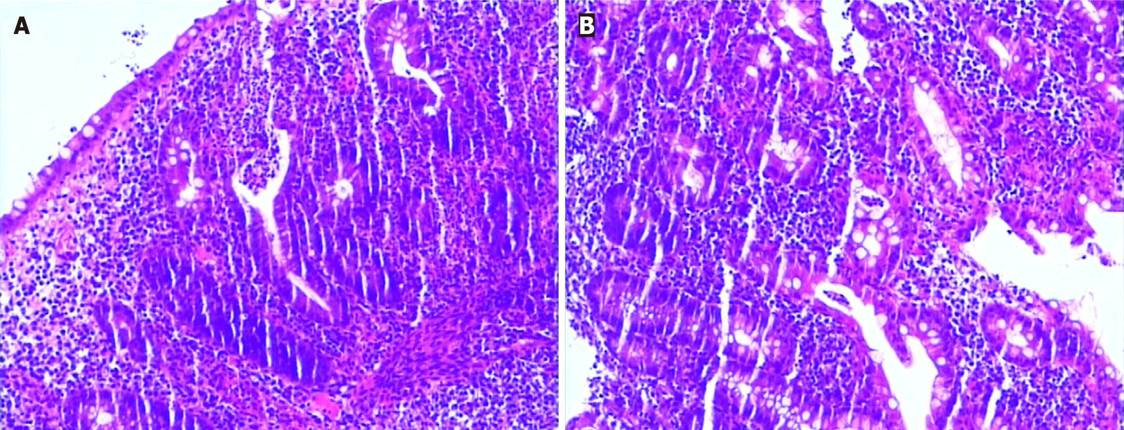
Figure 2 Findings of sigmoid colon biopsy specimens.
A: Histopathological findings of specimens [hematoxylin and eosin (HE), × 40)] with diffuse lymphocytic infiltration, irregular surface epithelium; B: Histopathological findings of specimens (HE, × 40) with diffuse lymphocytic infiltration, visible cryptitis, a crypt abscess, and distorted crypt structure.
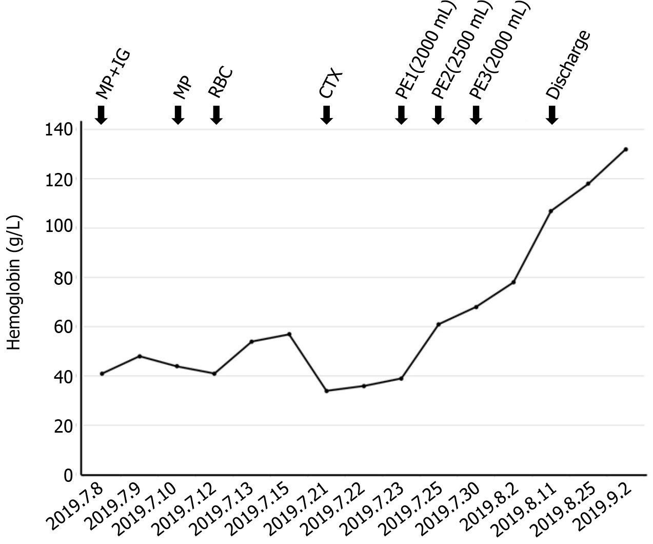
Figure 3 Clinical response to therapies.
MP: Methylprednisolone; IG: Immunoglobulin; RBC: Red blood cell; CTX: Cyclophosphamide; PE: Plasma exchange.
- Citation: Chen DX, Wu Y, Zhang SF, Yang XJ. Refractory autoimmune hemolytic anemia in a patient with systemic lupus erythematosus and ulcerative colitis: A case report. World J Clin Cases 2024; 12(13): 2286-2292
- URL: https://www.wjgnet.com/2307-8960/full/v12/i13/2286.htm
- DOI: https://dx.doi.org/10.12998/wjcc.v12.i13.2286


