Copyright
©The Author(s) 2024.
World J Clin Cases. May 6, 2024; 12(13): 2237-2242
Published online May 6, 2024. doi: 10.12998/wjcc.v12.i13.2237
Published online May 6, 2024. doi: 10.12998/wjcc.v12.i13.2237
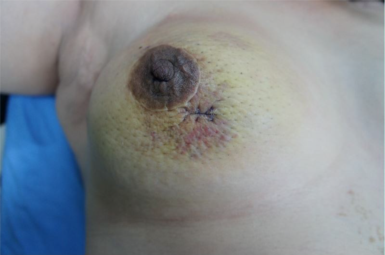
Figure 1 A skin ecchymotic plaque around the surgical scar on the right breast.
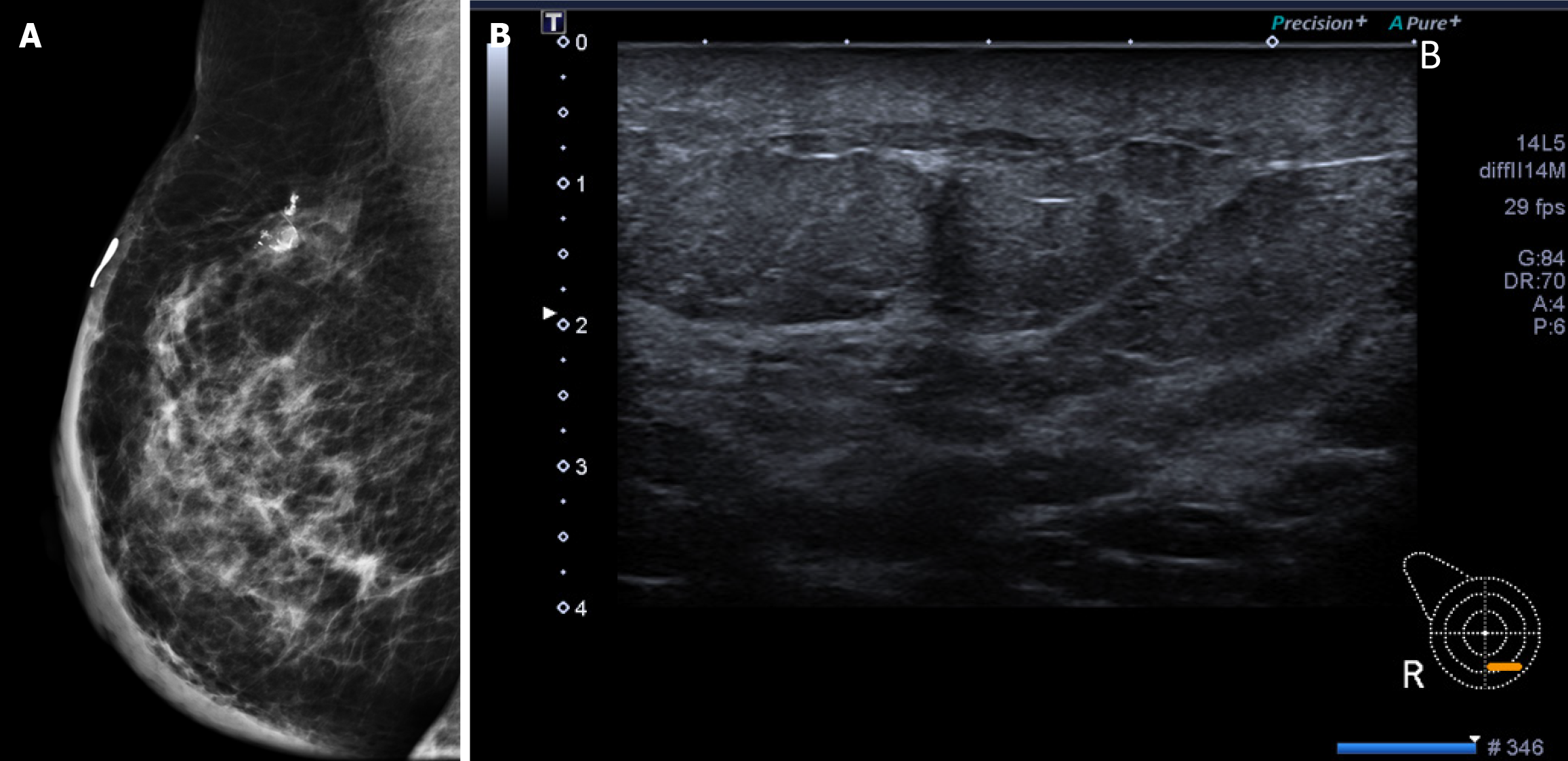
Figure 2 Mammography and ultrasound findings.
A: The mammography (medio-lateral oblique view) showed dystrophic calcifications in the upper hemisphere and diffuse skin thickening, misdiagnosed as post-operative changes initially; B: The B-mode ultrasound showed non-specific findings except skin thickenings.
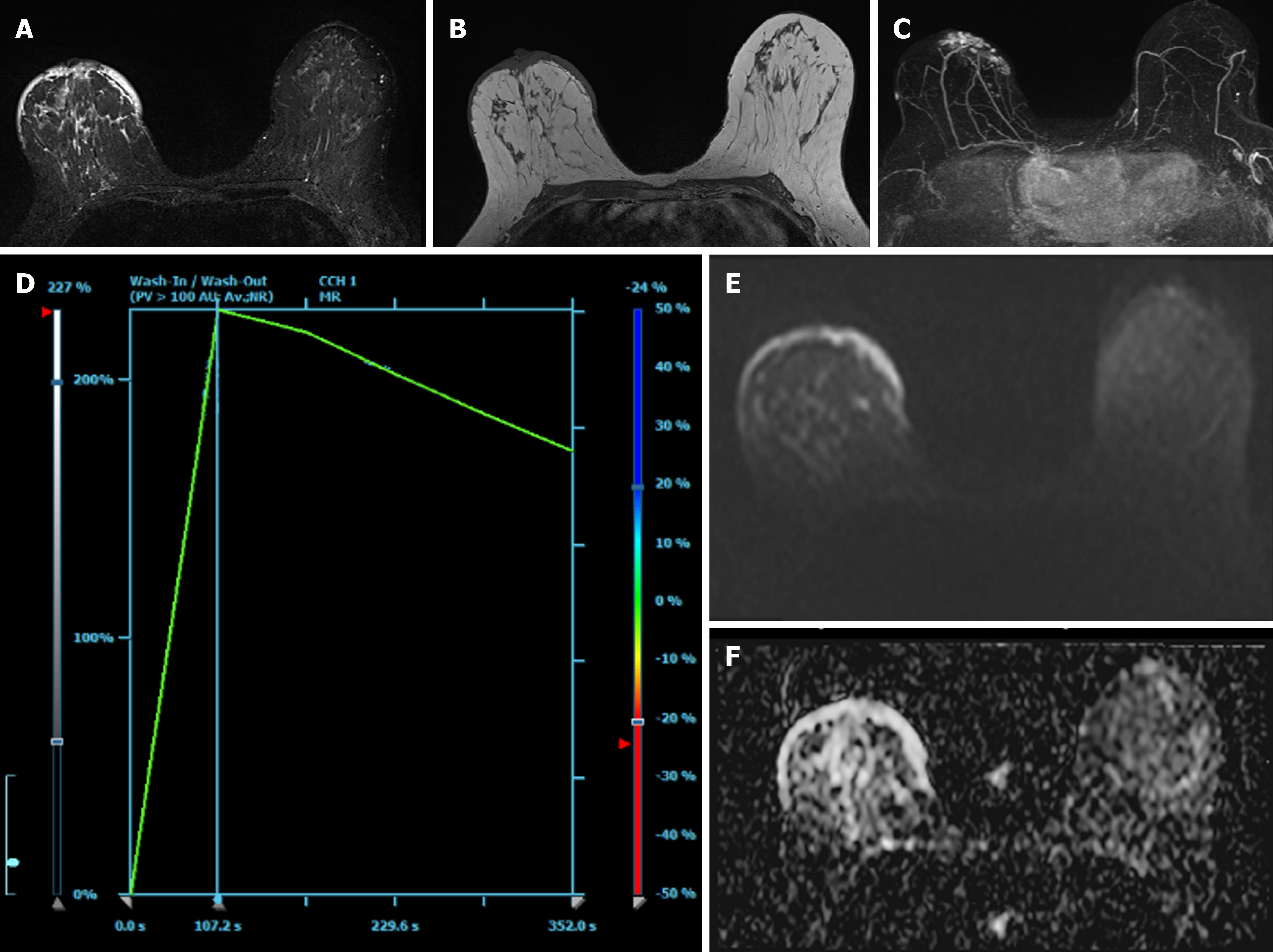
Figure 3 Illustrates the findings from contrast-enhanced breast magnetic resonance imaging, revealing diffuse skin thickening and nodularity.
A: Presurgical breast magnetic resonance imaging depicts diffuse skin thickening with hyperintensity on T2-weighted images; B: Diffuse skin thickening is observed on T1-weighted images; C: Diffuse skin thickening enhances on the maximum-intensity projection of postcontrast dynamic images; D: The enhancement kinetics of cutaneous lesions show fast wash-in and wash-out (type 3 curve); E: The axial diffusion-weighted image exhibits hyperintensity; F: The corresponding apparent diffusion coefficient map shows hyperintensity, indicating diffuse vasogenic edematous changes in the skin.
Figure 4 Case report timeline.
- Citation: Wu WP, Lee CW. Magnetic resonance imaging findings of radiation-induced breast angiosarcoma: A case report. World J Clin Cases 2024; 12(13): 2237-2242
- URL: https://www.wjgnet.com/2307-8960/full/v12/i13/2237.htm
- DOI: https://dx.doi.org/10.12998/wjcc.v12.i13.2237


