Copyright
©The Author(s) 2024.
World J Clin Cases. Apr 6, 2024; 12(10): 1810-1816
Published online Apr 6, 2024. doi: 10.12998/wjcc.v12.i10.1810
Published online Apr 6, 2024. doi: 10.12998/wjcc.v12.i10.1810
Figure 1 Colonoscopy findings.
A: Bluish purple mucosa with multiple ulcers in the ascending colon; B: A deep ulcer in the rectum.
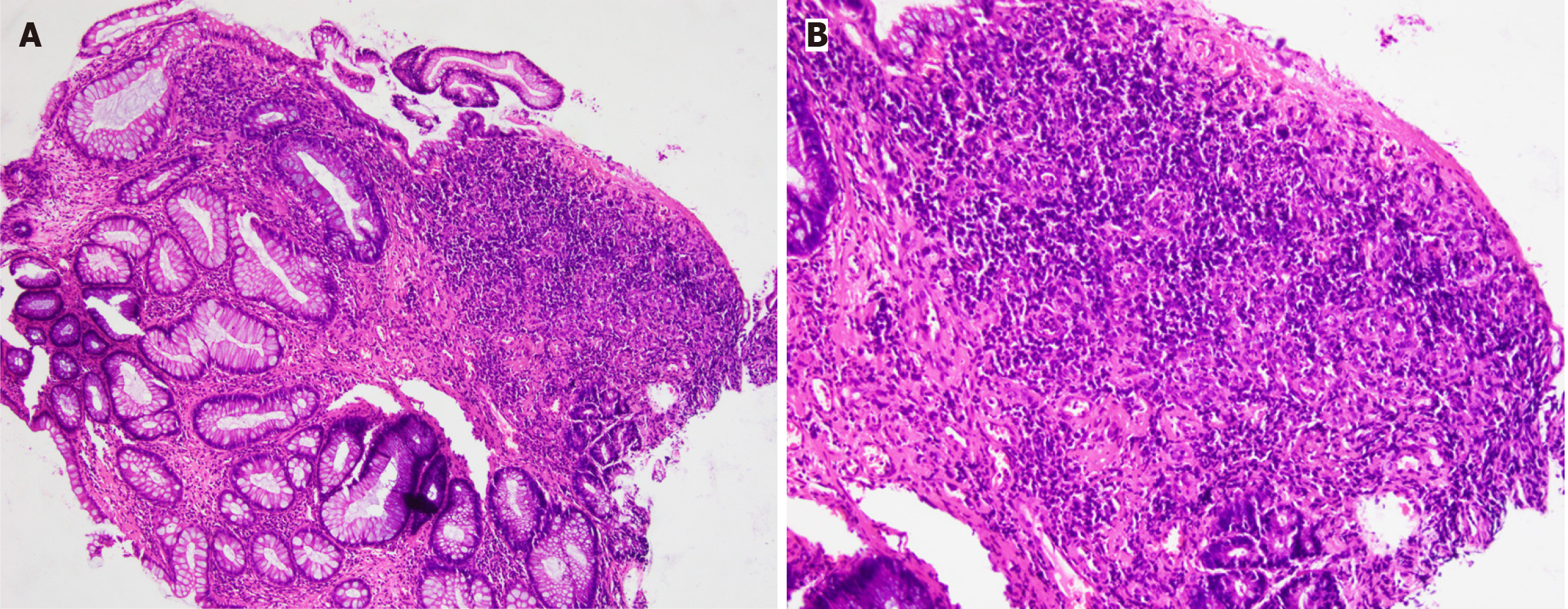
Figure 2 Histological examination.
A and B: Mild inflammation and granulomatous reaction were observed.
Figure 3 Plain abdominal computed tomography.
A-C: Diffuse mural thickening of the total colorectum and tortuous thread-like calcification were observed in the right hemicolon, left hemicolon, and rectum; D: Multiphase, contrast-enhanced computed tomography showed that most calcifications are located in the mesenteric vein.
Figure 4 Contrast-enhanced abdominal computed tomography.
A: Calcification of the abdominal aorta (arrow); B: Stratified calcification of the intestinal wall (arrow); C and D: The number of point intensifications in the arterial and venous phases in similar areas at the same level were seen to vary (box). There is more punctate reinforcement in the arterial phase than in the venous phase, and the punctate reinforcement in the venous phase is the calcification point.
- Citation: Wang M, Wan YX, Liao JW, Xiong F. Idiopathic mesenteric phlebosclerosis missed by a radiologist at initial diagnosis: A case report. World J Clin Cases 2024; 12(10): 1810-1816
- URL: https://www.wjgnet.com/2307-8960/full/v12/i10/1810.htm
- DOI: https://dx.doi.org/10.12998/wjcc.v12.i10.1810


