Copyright
©The Author(s) 2023.
World J Clin Cases. Mar 26, 2023; 11(9): 1951-1962
Published online Mar 26, 2023. doi: 10.12998/wjcc.v11.i9.1951
Published online Mar 26, 2023. doi: 10.12998/wjcc.v11.i9.1951
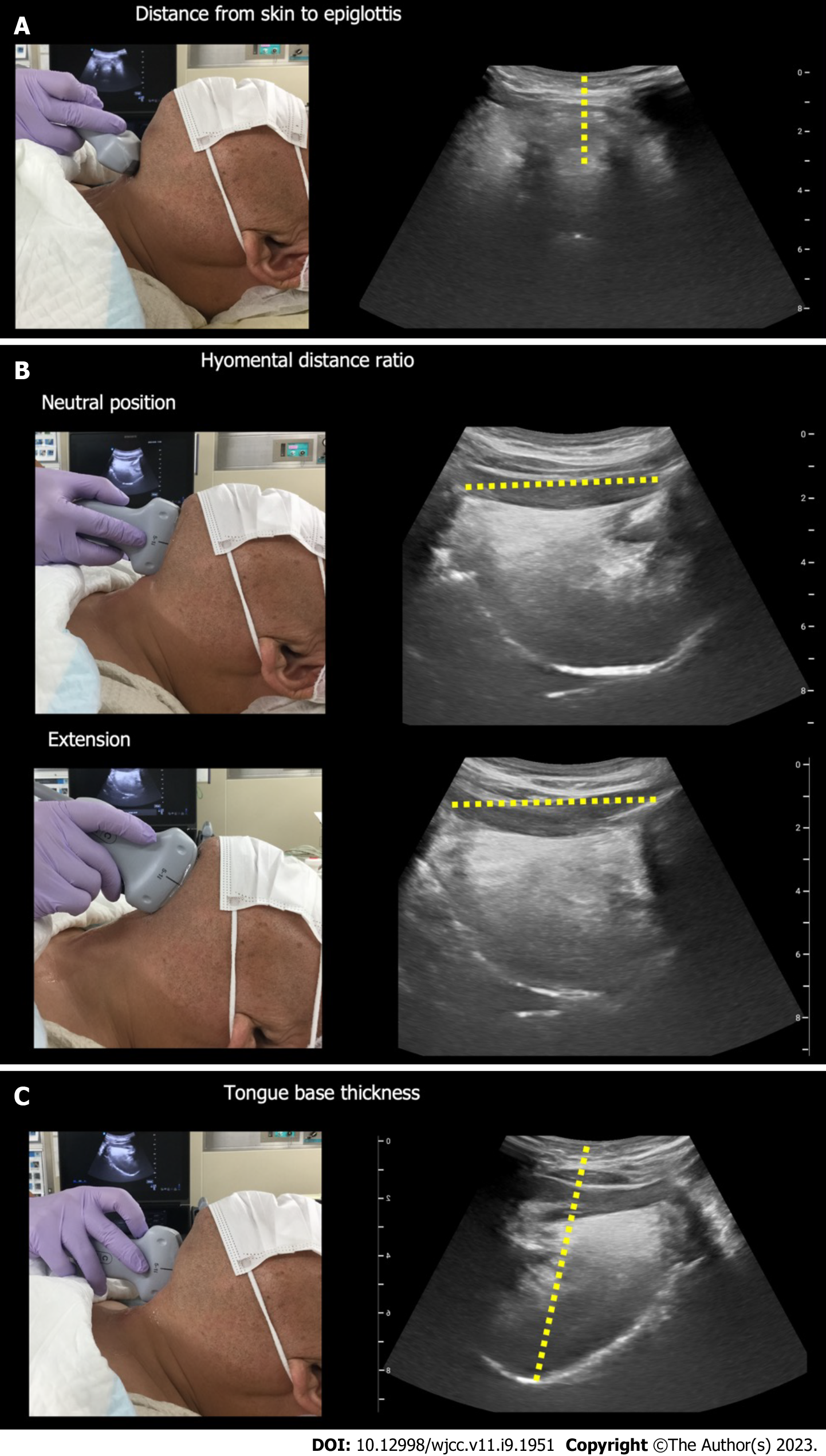
Figure 1 Predicting a difficult airway using airway ultrasound.
A: Ultrasound measurement of the distance from skin to epiglottis; B: Ultrasound measurement of the hyomental distance in the neutral and maximal extended neck positions; C: Ultrasound measurement of the distance from skin to tongue base.
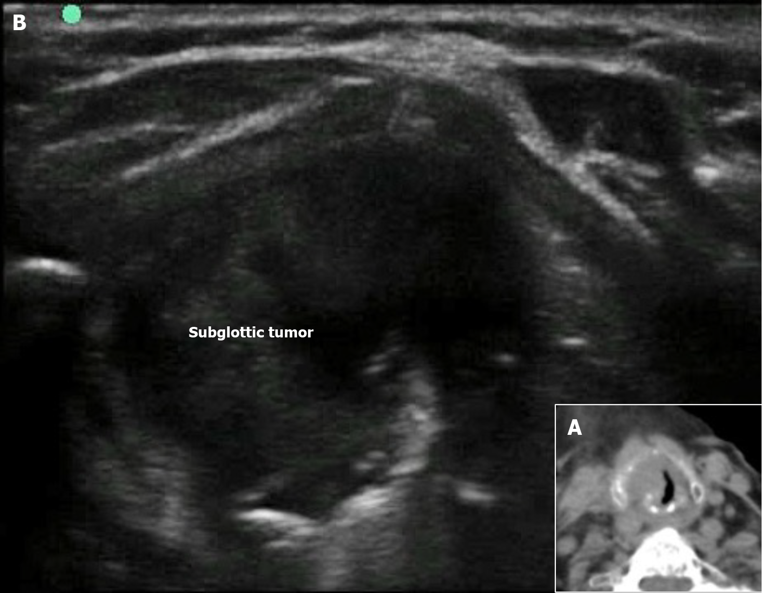
Figure 2 Subglottic tumor (reproduced from Figure 1 of Falcetta et al[22], with the permission of the copyright holder).
A: Transverse image from the computed tomography scan shows a subglottic mass; B: Ultrasound view at the level of the cricothyroid membrane shows a mass (subglottic tumor). Citation: Falcetta S, Cavallo S, Gabbanelli V, Pelaia P, Sorbello M, Zdravkovic I, Donati A. Evaluation of two neck ultrasound measurements as predictors of difficult direct laryngoscopy: A prospective observational study. Eur J Anaesthesiol 2018; 35: 605-612. Copyright© The Authors 2018. Published by Wolters Kluwer. The authors have obtained the permission for figure using (Supplementary material).
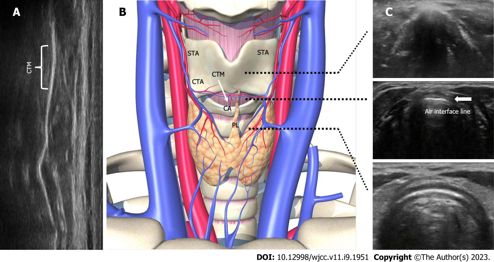
Figure 3 Anatomy and sonographic appearance of the cricothyroid membrane.
A: Longitudinal view for identifying the cricothyroid membrane; B: The thyroid, cricoid, and tracheal cartilages are strung together and appear like a string of pearls. Anatomy of the cricothyroid membrane; C: Transverse view for identifying the cricothyroid membrane. Air interface line indicates the cricothyroid membrane. CTM: Cricothyroid membrane; STA: Superior thyroid artery; CTA: Cricothyroid artery; CA: Common artery; PL: Pyramidal lobe of the thyroid gland.
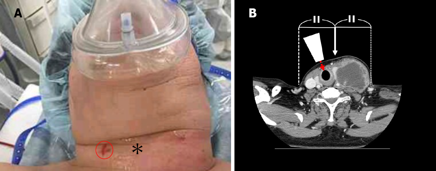
Figure 4 Identification of the cricothyroid membrane by airway ultrasound (reproduced from Figures 1 and 3 of Katayama et al[52], with the permission of the copyright holder).
A: The patient’s neck: The asterisk indicates the area palpated by the surgeon to find the cricothyroid membrane. The red circle shows the cricothyroid membrane identified by ultrasound; B: Cervical computed tomography scan image: The white arrow points to the apparent center of the neck. The true center (sagittal line) of the neck is shifted to the right. The ultrasound probe (white trapezoid) is placed perpendicularly to the skin and the ultrasound beam (red dashed arrow) is directed toward the cricothyroid membrane. Citation: Katayama A, Watanabe K, Tokumine J, Lefor AK, Nakazawa H, Jimbo I, Yorozu T. Cricothyroidotomy needle length is associated with posterior tracheal wall injury: A randomized crossover simulation study (CONSORT). Medicine (Baltimore) 2020; 99: e19331. Copyright© The Authors 2020. Published by MDPI. The authors have obtained the permission for figure using (Supplementary mate
- Citation: Nakazawa H, Uzawa K, Tokumine J, Lefor AK, Motoyasu A, Yorozu T. Airway ultrasound for patients anticipated to have a difficult airway: Perspective for personalized medicine. World J Clin Cases 2023; 11(9): 1951-1962
- URL: https://www.wjgnet.com/2307-8960/full/v11/i9/1951.htm
- DOI: https://dx.doi.org/10.12998/wjcc.v11.i9.1951


