Copyright
©The Author(s) 2023.
World J Clin Cases. Mar 16, 2023; 11(8): 1823-1829
Published online Mar 16, 2023. doi: 10.12998/wjcc.v11.i8.1823
Published online Mar 16, 2023. doi: 10.12998/wjcc.v11.i8.1823
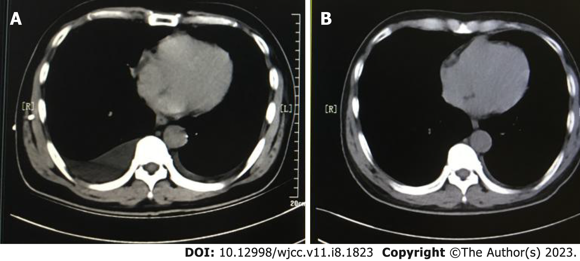
Figure 1 Patient's chest computed tomography on admission and at discharge.
A: Chest computed tomography showed bilateral pleural effusion; B: The effusion gradually resolved after 1 wk of treatment.
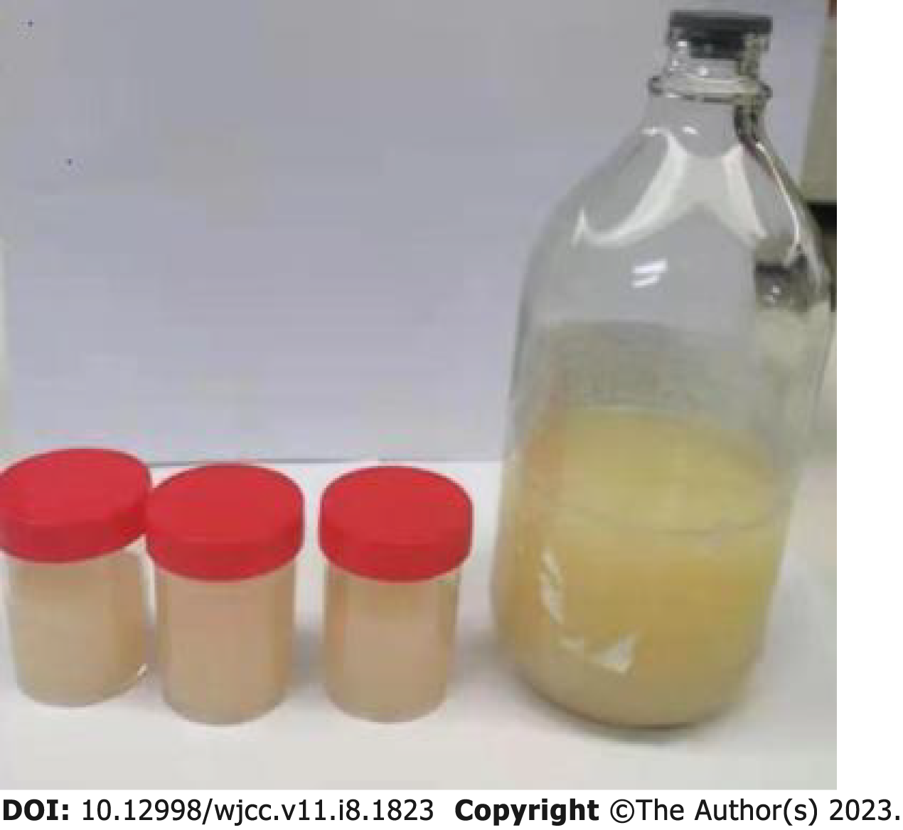
Figure 2 Fluid from the closed thoracic drainage.
The pleural effusion was milky white.
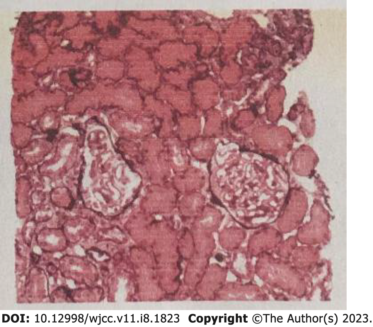
Figure 3 Renal biopsy results.
Biopsy revealed mild glomerular mesangium and stromal hyperplasia, focal segmental sclerosis, diffuse globular thickening of the basement membrane, diffuse segmental pegging, and unremarkable intracapillary hyperplasia. Mild tubular atrophy, extensive oedema, and unremarkable interstitial and vascular lesions were also present.
- Citation: Feng LL, Du J, Wang C, Wang SL. Primary membranous nephrotic syndrome with chylothorax as first presentation: A case report and literature review. World J Clin Cases 2023; 11(8): 1823-1829
- URL: https://www.wjgnet.com/2307-8960/full/v11/i8/1823.htm
- DOI: https://dx.doi.org/10.12998/wjcc.v11.i8.1823


