Copyright
©The Author(s) 2023.
World J Clin Cases. Jan 26, 2023; 11(3): 669-676
Published online Jan 26, 2023. doi: 10.12998/wjcc.v11.i3.669
Published online Jan 26, 2023. doi: 10.12998/wjcc.v11.i3.669
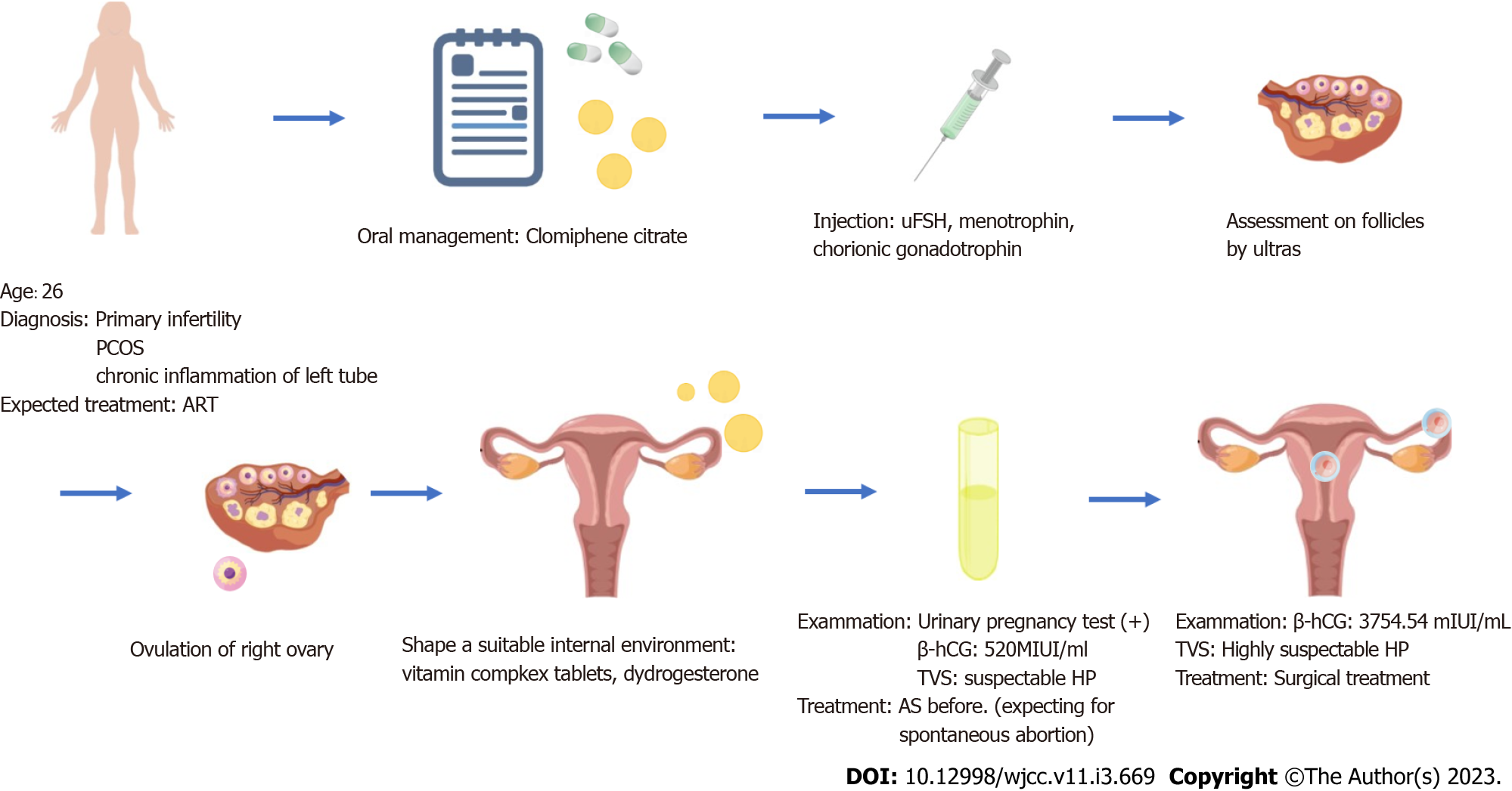
Figure 1 The complete process of diagnosis and treatment of the patient.
ART: Assisted reproductive techniques; hCG: Human chorionic gonadotropin; HP: Heterotopic pregnancy; PCOS: Polycystic ovary syndrome; TVS: Transvaginal scanning; uFSH: Urofollitropin for injection.
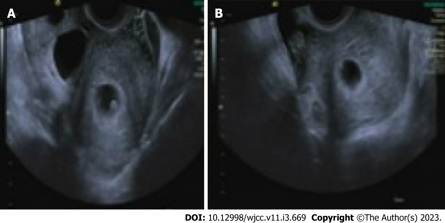
Figure 2 Transvaginal sonography.
A: The intrauterine gestational sac was about 7 wk of pregnancy; B: The bilateral ovaries were larger than normal. The left ovary measured 8 cm × 4 cm, while the right ovarian mass measured about 11 cm × 5 cm, suggestive of an ectopic pregnancy.
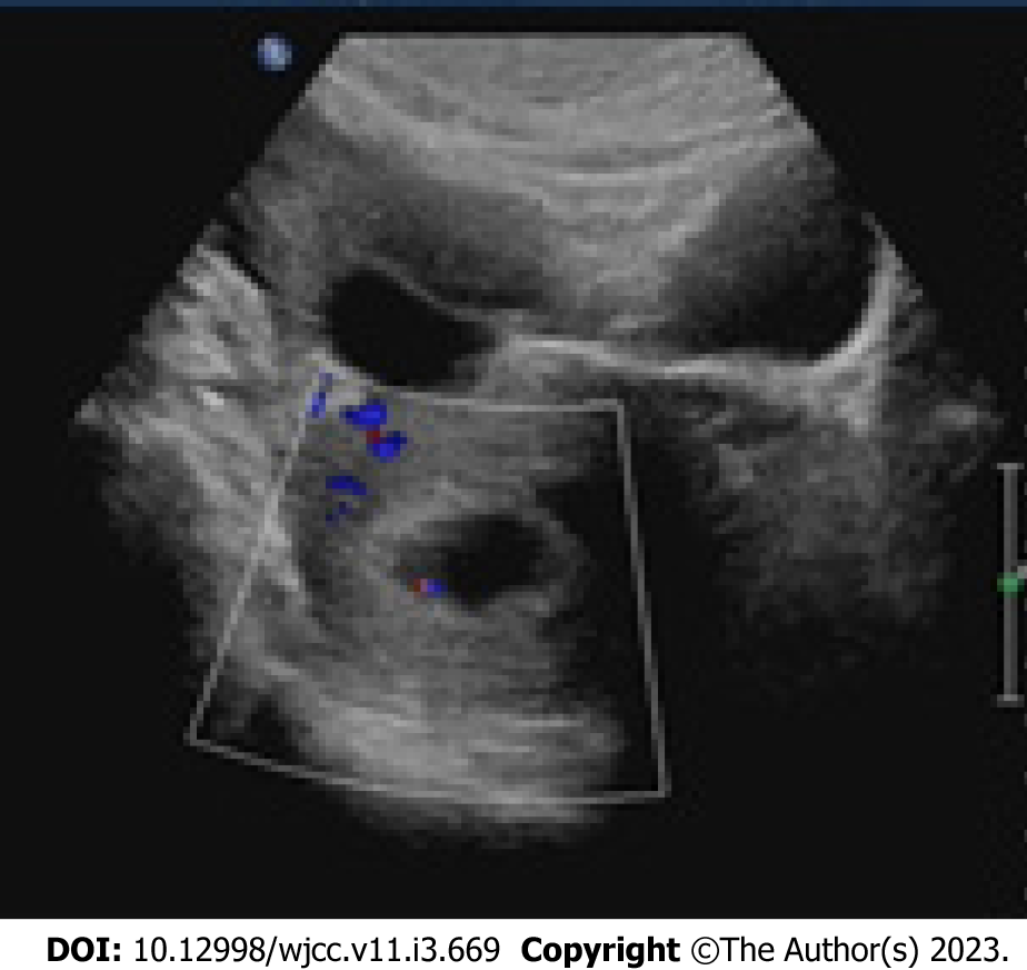
Figure 3
Re-examination by transvaginal sonography showing the intact intrauterine gestational sac.
Figure 4
Commonly used fertility treatments.
- Citation: Wang YN, Zheng LW, Fu LL, Xu Y, Zhang XY. Heterotopic pregnancy after assisted reproductive techniques with favorable outcome of the intrauterine pregnancy: A case report. World J Clin Cases 2023; 11(3): 669-676
- URL: https://www.wjgnet.com/2307-8960/full/v11/i3/669.htm
- DOI: https://dx.doi.org/10.12998/wjcc.v11.i3.669


