Copyright
©The Author(s) 2023.
World J Clin Cases. Jan 26, 2023; 11(3): 635-644
Published online Jan 26, 2023. doi: 10.12998/wjcc.v11.i3.635
Published online Jan 26, 2023. doi: 10.12998/wjcc.v11.i3.635
Figure 1 Initial intraoral examination of Case 1.
A: Tooth 21 was missing, and the alveolar nest was empty; B: There was no obvious lacerated wound in the gums; C: The root of the avulsed tooth was wiped clean, and no residual periodontal ligament tissue was observed.
Figure 2 Initial intraoral examination of Case 2.
A: Tooth 22 was missing, and the alveolar nest was empty; B: The patient had a complicated crown fracture of tooth 11 and a complicated crown-root fracture of tooth 21; C: The avulsed tooth was wrapped in dry paper towels, and obvious contaminants were present on the root surface.
Figure 3 Periapical radiograph and cone beam computed tomography images of Case 1 at the first visit.
A: The periodontal membrane space was widened in tooth 11, the alveolar socket of tooth 21 was empty, and no high-density foreign bodies were observed; B: Cone-beam computed tomography images showed that tooth 21 had been completely replanted, and the lip side of the alveolar bone wall of teeth 11 and 21 was fractured.
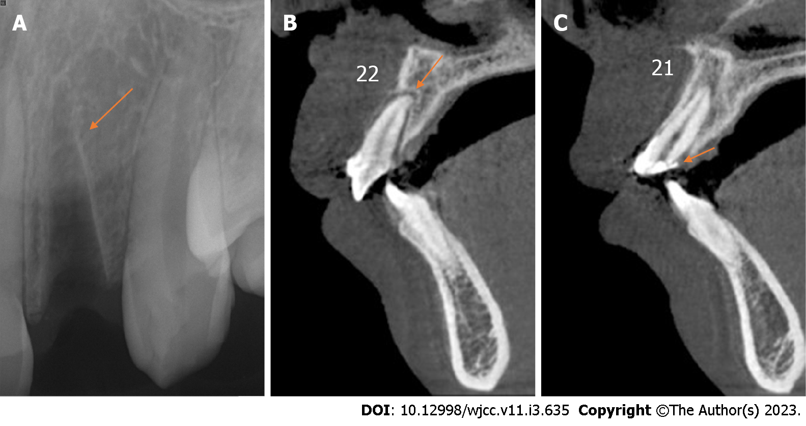
Figure 4 Periapical radiograph and cone beam computed tomography images of Case 2 at the first visit.
A: The alveolar socket of tooth 22 was empty, and no high-density foreign bodies were present; B: Cone-beam computed tomography image showed that tooth 22 had been replanted completely, and no alveolar fractures were noted around the empty tooth socket; C: Cone-beam computed tomography image showed that the fracture edge of tooth 21 was approximately 3 mm above the top of the alveolar crest.
Figure 5 Preparation process of the platelet-rich fibrin.
A: The tube was immediately centrifuged at 400 × g for 10 min; B: The fibrin clot contained platelet-rich fibrin and was located in the middle of the tube; C: The clot was easily separated from the red corpuscles at the bottom; D: The platelet-rich fibrin membrane was cut into approximately 1 mm3 granules, and the red and white ends were mixed evenly.
Figure 6 Intraoral photographs of replanted tooth fixation.
A and C: In Cases 1 and 2, the teeth were splinted using a preshaped semiflexible titanium labial arch; B and D: In Cases 1 and 2, the fixtures were removed.
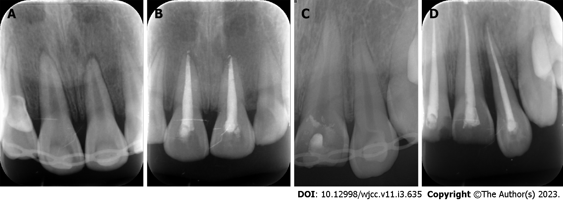
Figure 7 Periapical radiograph.
A and C: In Case 1 and 2, the teeth were splinted using a preshaped semiflexible titanium labial arch at the first visit; B and D: 6 wk after the initial visit of Case 1 and 2, the fixtures were removed, and the root canal treatment for the injured teeth was accomplished.
Figure 8 Intraoral photographs of the follow-up examination.
A and B: There were no obvious periodontal bags, tooth discoloration or swelling of the gums around tooth 21 in Case 1; C and D: There were no obvious periodontal bags, tooth discoloration or swelling of the gums around tooth 22 in Case 2.
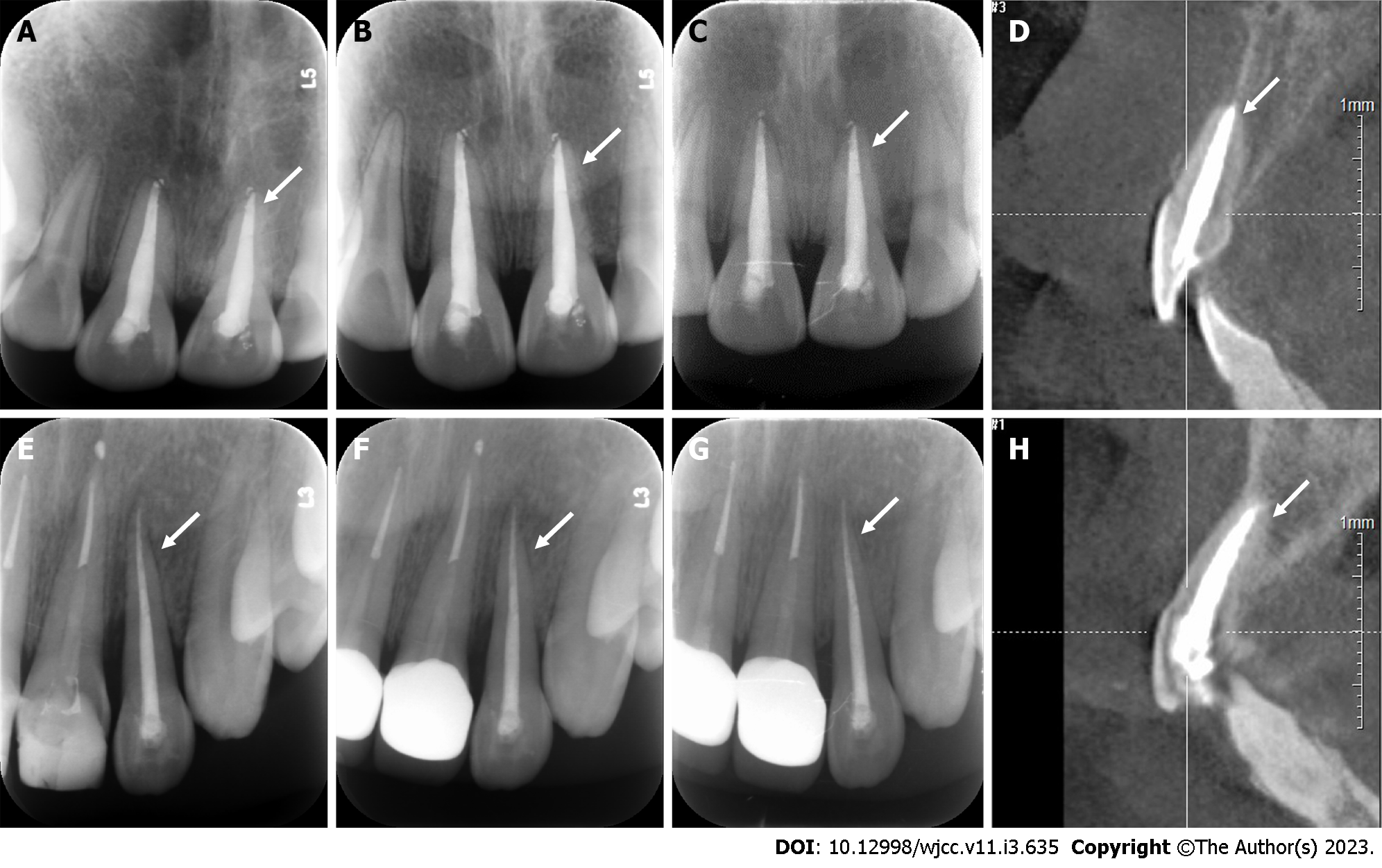
Figure 9 Periapical radiograph and cone beam computed tomography images of two cases during follow-up observation.
The periodontal membrane space was continuous, and no sign of pathological root absorption was observed in the avulsed teeth. A and E: 3-mo follow-up radiograph images of Cases 1 and 2; B and F: 6-mo follow-up radiograph images of Cases 1 and 2; C and G: 12-mo follow-up radiograph of Cases 1 and 2; D and H: 12-mo follow-up cone-beam computed tomography images of Cases 1 and 2.
- Citation: Yang Y, Liu YL, Jia LN, Wang JJ, Zhang M. Rescuing “hopeless” avulsed teeth using autologous platelet-rich fibrin following delayed reimplantation: Two case reports. World J Clin Cases 2023; 11(3): 635-644
- URL: https://www.wjgnet.com/2307-8960/full/v11/i3/635.htm
- DOI: https://dx.doi.org/10.12998/wjcc.v11.i3.635


