Copyright
©The Author(s) 2023.
World J Clin Cases. Oct 16, 2023; 11(29): 7200-7206
Published online Oct 16, 2023. doi: 10.12998/wjcc.v11.i29.7200
Published online Oct 16, 2023. doi: 10.12998/wjcc.v11.i29.7200
Figure 1 Gene sequencing.
A: Gene sequencing of the mother of the patient; B: Gene sequencing of older brother of the patient.
Figure 2 Bone marrow cytology and bone marrow biopsy.
A: Substantial active myeloid proliferation, with 14 granular megakaryocytes, one platelet-producing megakaryocyte, one naked megakaryocyte, and rare scattered platelets; B: Active nucleated cell proliferation, with 16 megakaryocytes and one plate-producing megakaryocyte; C: Active nucleated cell proliferation with 10 megakaryocytes and two plate-producing megakaryocytes; D: An increased proportion of lymphocytes and neutral-lobulated nucleated cells; E: Active nucleated cell proliferation, with 25 megakaryocytes and one plate-producing megakaryocyte; F: Extremely low bone marrow proliferation and lack of hematopoietic cells; G: Inhomogeneous proliferation of the bone marrow hematopoietic tissues; H: Low bone marrow hypoproliferation. I: Mild bone marrow hyperplasia; J: Active nucleated cell proliferation, triple-lineage cell differentiation and maturation.
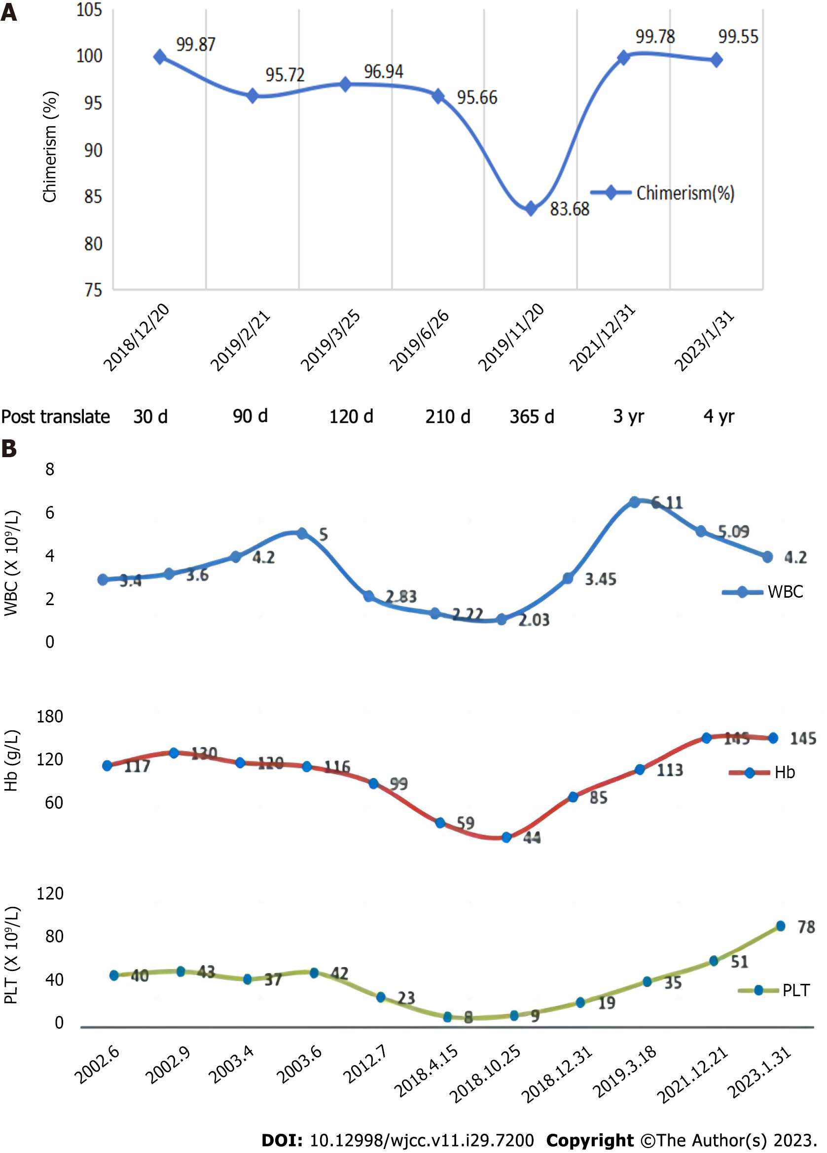
Figure 3 Bone marrow chimerism and blood count.
A: Bone marrow chimerism of the patient; B: Blood count of the patient.
Figure 4 Telomere length of the patient and his older brother.
A: The telomere length of the patient was distributed between 2.40 kb and 11.90 kb, and the mean telomere length was 6.01 kb, which was significantly shorter compared to age-matched healthy controls; B: The telomere length of the older brother was distributed between 3.31 kb and 12.46 kb, and the mean telomere length was 6.35 kb, which was normal when compared to age-matched healthy controls.
- Citation: Yan J, Jin T, Wang L. Hematopoietic stem cell transplantation of aplastic anemia by relative with mutations and normal telomere length: A case report. World J Clin Cases 2023; 11(29): 7200-7206
- URL: https://www.wjgnet.com/2307-8960/full/v11/i29/7200.htm
- DOI: https://dx.doi.org/10.12998/wjcc.v11.i29.7200


